5UN1

Crystal structure of GluN1/GluN2B delta-ATD NMDA receptor


Experimental Data Snapshot

  • Method: X-RAY DIFFRACTION
  • Resolution: 3.60 Å
  • R-Value Free: 0.316 
  • R-Value Work: 0.288 
  • R-Value Observed: 0.289 

wwPDB Validation   3D Report Full Report


Ligand Structure Quality Assessment 


This is version 1.5 of the entry. See complete history


Literature

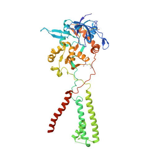
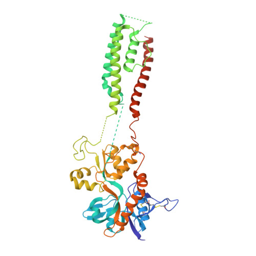
Mechanism of NMDA receptor channel block by MK-801 and memantine.

Song, X.Jensen, M.O.Jogini, V.Stein, R.A.Lee, C.H.Mchaourab, H.S.Shaw, D.E.Gouaux, E.

(2018) Nature 556: 515-519

  • DOI: https://doi.org/10.1038/s41586-018-0039-9
  • Primary Citation of Related Structures:  
    5UN1

  • PubMed Abstract: 

    The NMDA (N-methyl-D-aspartate) receptor transduces the binding of glutamate and glycine, coupling it to the opening of a calcium-permeable ion channel 1 . Owing to the lack of high-resolution structural studies of the NMDA receptor, the mechanism by which ion-channel blockers occlude ion permeation is not well understood. Here we show that removal of the amino-terminal domains from the GluN1-GluN2B NMDA receptor yields a functional receptor and crystals with good diffraction properties, allowing us to map the binding site of the NMDA receptor blocker, MK-801. This crystal structure, together with long-timescale molecular dynamics simulations, shows how MK-801 and memantine (a drug approved for the treatment of Alzheimer's disease) bind within the vestibule of the ion channel, promote closure of the ion channel gate and lodge between the M3-helix-bundle crossing and the M2-pore loops, physically blocking ion permeation.


  • Organizational Affiliation

    Vollum Institute, Oregon Health & Science University, Portland, OR, USA.


Macromolecules
Find similar proteins by:  (by identity cutoff)  |  3D Structure
Entity ID: 1
MoleculeChains Sequence LengthOrganismDetailsImage
N-methyl-D-aspartate receptor subunit NR1-3aA [auth G],
B [auth A],
E,
G [auth C]
451Xenopus laevisMutation(s): 0 
Membrane Entity: Yes 
UniProt
Find proteins for A0A1L8F5J9 (Xenopus laevis)
Explore A0A1L8F5J9 
Go to UniProtKB:  A0A1L8F5J9
Entity Groups  
Sequence Clusters30% Identity50% Identity70% Identity90% Identity95% Identity100% Identity
UniProt GroupA0A1L8F5J9
Sequence Annotations
Expand
  • Reference Sequence
Find similar proteins by:  (by identity cutoff)  |  3D Structure
Entity ID: 2
MoleculeChains Sequence LengthOrganismDetailsImage
Ionotropic glutamate receptor subunit NR2BC [auth B],
D [auth H],
F,
H [auth D]
448Xenopus laevisMutation(s): 0 
Gene Names: NR2BXELAEV_18025529mg
Membrane Entity: Yes 
UniProt
Find proteins for A7XY94 (Xenopus laevis)
Explore A7XY94 
Go to UniProtKB:  A7XY94
Entity Groups  
Sequence Clusters30% Identity50% Identity70% Identity90% Identity95% Identity100% Identity
UniProt GroupA7XY94
Sequence Annotations
Expand
  • Reference Sequence
Small Molecules
Ligands 7 Unique
IDChains Name / Formula / InChI Key2D Diagram3D Interactions
Y01
Query on Y01

Download Ideal Coordinates CCD File 
V [auth E]CHOLESTEROL HEMISUCCINATE
C31 H50 O4
WLNARFZDISHUGS-MIXBDBMTSA-N
BMK
Query on BMK

Download Ideal Coordinates CCD File 
M [auth G],
S [auth B]
(5S,10R)-5-methyl-10,11-dihydro-5H-5,10-epiminodibenzo[a,d][7]annulene
C16 H15 N
LBOJYSIDWZQNJS-CVEARBPZSA-N
NAG
Query on NAG

Download Ideal Coordinates CCD File 
J [auth G]
R [auth B]
U [auth H]
W [auth F]
X [auth D]
J [auth G],
R [auth B],
U [auth H],
W [auth F],
X [auth D],
Z [auth D]
2-acetamido-2-deoxy-beta-D-glucopyranose
C8 H15 N O6
OVRNDRQMDRJTHS-FMDGEEDCSA-N
GLU
Query on GLU

Download Ideal Coordinates CCD File 
L [auth G],
N [auth G],
Q [auth B],
T [auth H],
Y [auth D]
GLUTAMIC ACID
C5 H9 N O4
WHUUTDBJXJRKMK-VKHMYHEASA-N
TRS
Query on TRS

Download Ideal Coordinates CCD File 
K [auth G]2-AMINO-2-HYDROXYMETHYL-PROPANE-1,3-DIOL
C4 H12 N O3
LENZDBCJOHFCAS-UHFFFAOYSA-O
GLY
Query on GLY

Download Ideal Coordinates CCD File 
I [auth G],
P [auth A]
GLYCINE
C2 H5 N O2
DHMQDGOQFOQNFH-UHFFFAOYSA-N
MG
Query on MG

Download Ideal Coordinates CCD File 
O [auth A]MAGNESIUM ION
Mg
JLVVSXFLKOJNIY-UHFFFAOYSA-N
Binding Affinity Annotations 
IDSourceBinding Affinity
BMK BindingDB:  5UN1 Ki: min: 8, max: 31 (nM) from 3 assay(s)
IC50: 29 (nM) from 1 assay(s)
Experimental Data & Validation

Experimental Data

  • Method: X-RAY DIFFRACTION
  • Resolution: 3.60 Å
  • R-Value Free: 0.316 
  • R-Value Work: 0.288 
  • R-Value Observed: 0.289 
  • Space Group: P 1 21 1
Unit Cell:
Length ( Å )Angle ( ˚ )
a = 181.57α = 90
b = 108.47β = 111.44
c = 182.46γ = 90
Software Package:
Software NamePurpose
PHENIXrefinement
XDSdata reduction
Aimlessdata scaling
PHASERphasing

Structure Validation

View Full Validation Report


Ligand Structure Quality Assessment 


Entry History & Funding Information

Deposition Data


Funding OrganizationLocationGrant Number
National Institutes of Health/National Institute of Neurological Disorders and Stroke (NIH/NINDS)United StatesR01-NS038631

Revision History  (Full details and data files)

  • Version 1.0: 2018-02-07
    Type: Initial release
  • Version 1.1: 2018-04-18
    Changes: Data collection, Database references
  • Version 1.2: 2018-05-02
    Changes: Data collection, Database references
  • Version 1.3: 2020-07-29
    Type: Remediation
    Reason: Carbohydrate remediation
    Changes: Data collection, Derived calculations, Structure summary
  • Version 1.4: 2022-04-13
    Changes: Author supporting evidence, Database references, Structure summary
  • Version 1.5: 2023-10-04
    Changes: Data collection, Refinement description