Coordination chemistry controls the thiol oxidase activity of the B12-trafficking protein CblC.
Li, Z., Shanmuganathan, A., Ruetz, M., Yamada, K., Lesniak, N.A., Krautler, B., Brunold, T.C., Koutmos, M., Banerjee, R.(2017) J Biological Chem 292: 9733-9744
- PubMed: 28442570 Search on PubMedSearch on PubMed Central
- DOI: https://doi.org/10.1074/jbc.M117.788554
- Primary Citation Related Structures:
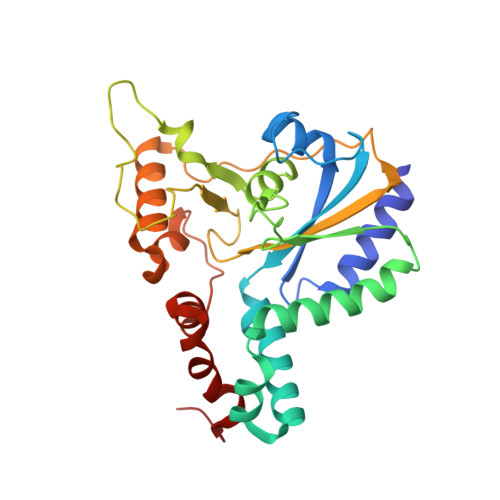
5UJC - PubMed Abstract:
The cobalamin or B 12 cofactor supports sulfur and one-carbon metabolism and the catabolism of odd-chain fatty acids, branched-chain amino acids, and cholesterol. CblC is a B 12 -processing enzyme involved in an early cytoplasmic step in the cofactor-trafficking pathway. It catalyzes the glutathione (GSH)-dependent dealkylation of alkylcobalamins and the reductive decyanation of cyanocobalamin. CblC from Caenorhabditis elegans ( ce CblC) also exhibits a robust thiol oxidase activity, converting reduced GSH to oxidized GSSG with concomitant scrubbing of ambient dissolved O 2 The mechanism of thiol oxidation catalyzed by ce CblC is not known. In this study, we demonstrate that novel coordination chemistry accessible to ce CblC-bound cobalamin supports its thiol oxidase activity via a glutathionyl-cobalamin intermediate. Deglutathionylation of glutathionyl-cobalamin by a second molecule of GSH yields GSSG. The crystal structure of ce CblC provides insights into how architectural differences at the α- and β-faces of cobalamin promote the thiol oxidase activity of ce CblC but mute it in wild-type human CblC. The R161G and R161Q mutations in human CblC unmask its latent thiol oxidase activity and are correlated with increased cellular oxidative stress disease. In summary, we have uncovered key architectural features in the cobalamin-binding pocket that support unusual cob(II)alamin coordination chemistry and enable the thiol oxidase activity of ce CblC.
- From the Department of Biological Chemistry, University of Michigan Medical Center, Ann Arbor, Michigan 48109-0600.
Organizational Affiliation: