5UFE | pdb_00005ufe

Wild-type K-Ras(GNP)/R11.1.6 complex


Experimental Data Snapshot

  • Method: X-RAY DIFFRACTION
  • Resolution: 2.30 Å
  • R-Value Free: 
    0.241 (Depositor), 0.241 (DCC) 
  • R-Value Work: 
    0.184 (Depositor), 0.186 (DCC) 
  • R-Value Observed: 
    0.190 (Depositor) 

Starting Models: experimental
View more details

wwPDB Validation 3D Report Full Report

Validation slider image for 5UFE

Ligand Structure Quality Assessment 


This is version 1.3 of the entry. See complete history

Literature

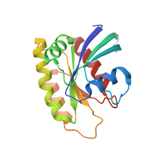
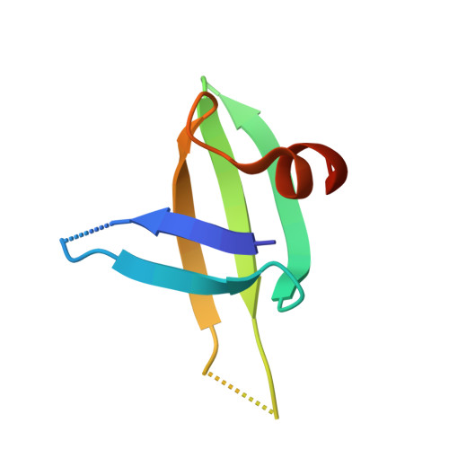
An engineered protein antagonist of K-Ras/B-Raf interaction.

Kauke, M.J.Traxlmayr, M.W.Parker, J.A.Kiefer, J.D.Knihtila, R.McGee, J.Verdine, G.Mattos, C.Wittrup, K.D.

(2017) Sci Rep 7: 5831-5831

  • DOI: https://doi.org/10.1038/s41598-017-05889-7
  • Primary Citation Related Structures: 
    5UFE, 5UFQ

  • PubMed Abstract: 

    Ras is at the hub of signal transduction pathways controlling cell proliferation and survival. Its mutants, present in about 30% of human cancers, are major drivers of oncogenesis and render tumors unresponsive to standard therapies. Here we report the engineering of a protein scaffold for preferential binding to K-Ras G12D. This is the first reported inhibitor to achieve nanomolar affinity while exhibiting specificity for mutant over wild type (WT) K-Ras. Crystal structures of the protein R11.1.6 in complex with K-Ras WT and K-Ras G12D offer insight into the structural basis for specificity, highlighting differences in the switch I conformation as the major defining element in the higher affinity interaction. R11.1.6 directly blocks interaction with Raf and reduces signaling through the Raf/MEK/ERK pathway. Our results support greater consideration of the state of switch I and provide a novel tool to study Ras biology. Most importantly, this work makes an unprecedented contribution to Ras research in inhibitor development strategy by revealing details of a targetable binding surface. Unlike the polar interfaces found for Ras/effector interactions, the K-Ras/R11.1.6 complex reveals an extensive hydrophobic interface that can serve as a template to advance the development of high affinity, non-covalent inhibitors of K-Ras oncogenic mutants.


  • Organizational Affiliation
    • Department of Chemical Engineering, Massachusetts Institute of Technology, Cambridge, MA, 02139, USA.

Macromolecule Content 

  • Total Structure Weight: 27.54 kDa 
  • Atom Count: 1,874 
  • Modeled Residue Count: 220 
  • Deposited Residue Count: 227 
  • Unique protein chains: 2

Macromolecules

Find similar proteins by:|  3D Structure
Entity ID: 1
MoleculeChains  Sequence LengthOrganismDetailsImage
GTPase KRas166Homo sapiensMutation(s): 0 
Gene Names: KRASKRAS2RASK2
EC: 3.6.5.2
UniProt & NIH Common Fund Data Resources
Find proteins for P01116 (Homo sapiens)
Explore P01116 
Go to UniProtKB:  P01116
PHAROS:  P01116
GTEx:  ENSG00000133703 
Entity Groups
Sequence Clusters30% Identity50% Identity70% Identity90% Identity95% Identity100% Identity
UniProt GroupP01116
Sequence Annotations
Expand
Reference Sequence
Find similar proteins by:|  3D Structure
Entity ID: 2
MoleculeChains  Sequence LengthOrganismDetailsImage
R11.1.661Saccharolobus solfataricusMutation(s): 0 

Small Molecules

Ligands 6 Unique
IDChains Name / Formula / InChI Key2D Diagram3D Interactions
GNP

Query on GNP


Download:Ideal Coordinates CCD File
C [auth A]PHOSPHOAMINOPHOSPHONIC ACID-GUANYLATE ESTER
C10 H17 N6 O13 P3
UQABYHGXWYXDTK-UUOKFMHZSA-N
CD

Query on CD


Download:Ideal Coordinates CCD File
F [auth A],
G [auth A],
H [auth A],
I [auth A]
CADMIUM ION
Cd
WLZRMCYVCSSEQC-UHFFFAOYSA-N
CO

Query on CO


Download:Ideal Coordinates CCD File
N [auth A],
O [auth A],
P [auth A]
COBALT (II) ION
Co
XLJKHNWPARRRJB-UHFFFAOYSA-N
CA

Query on CA


Download:Ideal Coordinates CCD File
D [auth A],
E [auth A],
Q [auth B]
CALCIUM ION
Ca
BHPQYMZQTOCNFJ-UHFFFAOYSA-N
CL

Query on CL


Download:Ideal Coordinates CCD File
J [auth A],
K [auth A],
L [auth A]
CHLORIDE ION
Cl
VEXZGXHMUGYJMC-UHFFFAOYSA-M
MG

Query on MG


Download:Ideal Coordinates CCD File
M [auth A]MAGNESIUM ION
Mg
JLVVSXFLKOJNIY-UHFFFAOYSA-N

Experimental Data & Validation

Experimental Data

  • Method: X-RAY DIFFRACTION
  • Resolution: 2.30 Å
  • R-Value Free:  0.241 (Depositor), 0.241 (DCC) 
  • R-Value Work:  0.184 (Depositor), 0.186 (DCC) 
  • R-Value Observed: 0.190 (Depositor) 
Space Group: P 21 21 21
Unit Cell:
Length ( Å )Angle ( ˚ )
a = 48.713α = 90
b = 64.648β = 90
c = 74.506γ = 90
Software Package:
Software NamePurpose
PHENIXrefinement
HKL-3000data reduction
HKL-3000data scaling
PHENIXphasing

Structure Validation

View Full Validation Report


Ligand Structure Quality Assessment 


Entry History 

& Funding Information

Deposition Data


Funding OrganizationLocationGrant Number
National Science Foundation (NSF, United States)United StatesMCB-1517295

Revision History  (Full details and data files)

  • Version 1.0: 2017-08-02
    Type: Initial release
  • Version 1.1: 2019-11-20
    Changes: Derived calculations
  • Version 1.2: 2019-11-27
    Changes: Author supporting evidence
  • Version 1.3: 2023-10-04
    Changes: Data collection, Database references, Derived calculations, Refinement description