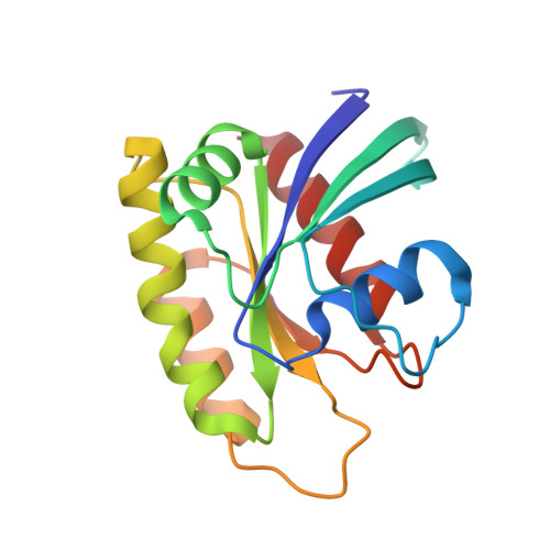
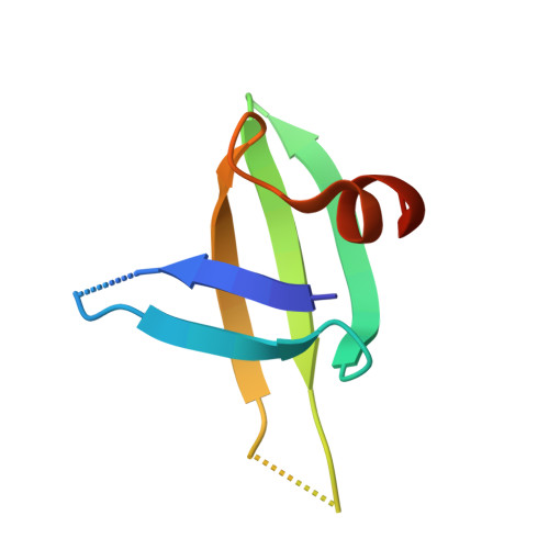
5UFE

Wild-type K-Ras(GNP)/R11.1.6 complex


Experimental Data Snapshot

  • Method: X-RAY DIFFRACTION
  • Resolution: 2.30 Å
  • R-Value Free: 0.241 
  • R-Value Work: 0.184 
  • R-Value Observed: 0.190 

Starting Models: experimental
View more details

wwPDB Validation   3D Report Full Report


Ligand Structure Quality Assessment 


This is version 1.3 of the entry. See complete history


Literature

An engineered protein antagonist of K-Ras/B-Raf interaction.

Kauke, M.J.Traxlmayr, M.W.Parker, J.A.Kiefer, J.D.Knihtila, R.McGee, J.Verdine, G.Mattos, C.Wittrup, K.D.

(2017) Sci Rep 7: 5831-5831

  • DOI: https://doi.org/10.1038/s41598-017-05889-7
  • Primary Citation of Related Structures:  
    5UFE, 5UFQ

  • PubMed Abstract: 

    Ras is at the hub of signal transduction pathways controlling cell proliferation and survival. Its mutants, present in about 30% of human cancers, are major drivers of oncogenesis and render tumors unresponsive to standard therapies. Here we report the engineering of a protein scaffold for preferential binding to K-Ras G12D. This is the first reported inhibitor to achieve nanomolar affinity while exhibiting specificity for mutant over wild type (WT) K-Ras. Crystal structures of the protein R11.1.6 in complex with K-Ras WT and K-Ras G12D offer insight into the structural basis for specificity, highlighting differences in the switch I conformation as the major defining element in the higher affinity interaction. R11.1.6 directly blocks interaction with Raf and reduces signaling through the Raf/MEK/ERK pathway. Our results support greater consideration of the state of switch I and provide a novel tool to study Ras biology. Most importantly, this work makes an unprecedented contribution to Ras research in inhibitor development strategy by revealing details of a targetable binding surface. Unlike the polar interfaces found for Ras/effector interactions, the K-Ras/R11.1.6 complex reveals an extensive hydrophobic interface that can serve as a template to advance the development of high affinity, non-covalent inhibitors of K-Ras oncogenic mutants.


  • Organizational Affiliation

    Department of Chemical Engineering, Massachusetts Institute of Technology, Cambridge, MA, 02139, USA.


Macromolecules
Find similar proteins by:  (by identity cutoff)  |  3D Structure
Entity ID: 1
MoleculeChains Sequence LengthOrganismDetailsImage
GTPase KRas166Homo sapiensMutation(s): 0 
Gene Names: KRASKRAS2RASK2
EC: 3.6.5.2
UniProt & NIH Common Fund Data Resources
Find proteins for P01116 (Homo sapiens)
Explore P01116 
Go to UniProtKB:  P01116
PHAROS:  P01116
GTEx:  ENSG00000133703 
Entity Groups  
Sequence Clusters30% Identity50% Identity70% Identity90% Identity95% Identity100% Identity
UniProt GroupP01116
Sequence Annotations
Expand
  • Reference Sequence
Find similar proteins by:  (by identity cutoff)  |  3D Structure
Entity ID: 2
MoleculeChains Sequence LengthOrganismDetailsImage
R11.1.661Saccharolobus solfataricusMutation(s): 0 
Entity Groups  
Sequence Clusters30% Identity50% Identity70% Identity90% Identity95% Identity100% Identity
Sequence Annotations
Expand
  • Reference Sequence
Small Molecules
Ligands 6 Unique
IDChains Name / Formula / InChI Key2D Diagram3D Interactions
GNP
Query on GNP

Download Ideal Coordinates CCD File 
C [auth A]PHOSPHOAMINOPHOSPHONIC ACID-GUANYLATE ESTER
C10 H17 N6 O13 P3
UQABYHGXWYXDTK-UUOKFMHZSA-N
CD
Query on CD

Download Ideal Coordinates CCD File 
F [auth A],
G [auth A],
H [auth A],
I [auth A]
CADMIUM ION
Cd
WLZRMCYVCSSEQC-UHFFFAOYSA-N
CO
Query on CO

Download Ideal Coordinates CCD File 
N [auth A],
O [auth A],
P [auth A]
COBALT (II) ION
Co
XLJKHNWPARRRJB-UHFFFAOYSA-N
CA
Query on CA

Download Ideal Coordinates CCD File 
D [auth A],
E [auth A],
Q [auth B]
CALCIUM ION
Ca
BHPQYMZQTOCNFJ-UHFFFAOYSA-N
CL
Query on CL

Download Ideal Coordinates CCD File 
J [auth A],
K [auth A],
L [auth A]
CHLORIDE ION
Cl
VEXZGXHMUGYJMC-UHFFFAOYSA-M
MG
Query on MG

Download Ideal Coordinates CCD File 
M [auth A]MAGNESIUM ION
Mg
JLVVSXFLKOJNIY-UHFFFAOYSA-N
Experimental Data & Validation

Experimental Data

  • Method: X-RAY DIFFRACTION
  • Resolution: 2.30 Å
  • R-Value Free: 0.241 
  • R-Value Work: 0.184 
  • R-Value Observed: 0.190 
  • Space Group: P 21 21 21
Unit Cell:
Length ( Å )Angle ( ˚ )
a = 48.713α = 90
b = 64.648β = 90
c = 74.506γ = 90
Software Package:
Software NamePurpose
PHENIXrefinement
HKL-3000data reduction
HKL-3000data scaling
PHENIXphasing

Structure Validation

View Full Validation Report


Ligand Structure Quality Assessment 


Entry History & Funding Information

Deposition Data


Funding OrganizationLocationGrant Number
National Science Foundation (NSF, United States)United StatesMCB-1517295

Revision History  (Full details and data files)

  • Version 1.0: 2017-08-02
    Type: Initial release
  • Version 1.1: 2019-11-20
    Changes: Derived calculations
  • Version 1.2: 2019-11-27
    Changes: Author supporting evidence
  • Version 1.3: 2023-10-04
    Changes: Data collection, Database references, Derived calculations, Refinement description