5U09

High-resolution crystal structure of the human CB1 cannabinoid receptor


Experimental Data Snapshot

  • Method: X-RAY DIFFRACTION
  • Resolution: 2.60 Å
  • R-Value Free: 0.238 
  • R-Value Work: 0.180 
  • R-Value Observed: 0.183 

wwPDB Validation   3D Report Full Report


Ligand Structure Quality Assessment 


This is version 1.2 of the entry. See complete history


Literature

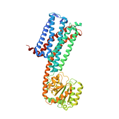
High-resolution crystal structure of the human CB1 cannabinoid receptor.

Shao, Z.Yin, J.Chapman, K.Grzemska, M.Clark, L.Wang, J.Rosenbaum, D.M.

(2016) Nature 

  • DOI: https://doi.org/10.1038/nature20613
  • Primary Citation of Related Structures:  
    5U09

  • PubMed Abstract: 

    The human cannabinoid G-protein-coupled receptors (GPCRs) CB1 and CB2 mediate the functional responses to the endocannabinoids anandamide and 2-arachidonyl glycerol (2-AG) and to the widely consumed plant phytocannabinoid Δ 9 -tetrahydrocannabinol (THC). The cannabinoid receptors have been the targets of intensive drug discovery efforts, because modulation of these receptors has therapeutic potential to control pain, epilepsy, obesity, and other disorders. Although much progress in understanding the biophysical properties of GPCRs has recently been made, investigations of the molecular mechanisms of the cannabinoids and their receptors have lacked high-resolution structural data. Here we report the use of GPCR engineering and lipidic cubic phase crystallization to determine the structure of the human CB1 receptor bound to the inhibitor taranabant at 2.6-Å resolution. We found that the extracellular surface of CB1, including the highly conserved membrane-proximal N-terminal region, is distinct from those of other lipid-activated GPCRs, forming a critical part of the ligand-binding pocket. Docking studies further demonstrate how this same pocket may accommodate the cannabinoid agonist THC. Our CB1 structure provides an atomic framework for studying cannabinoid receptor function and will aid the design and optimization of therapeutic modulators of the endocannabinoid system.


  • Organizational Affiliation

    Department of Biophysics, The University of Texas Southwestern Medical Center, Dallas, Texas 75390, USA.


Macromolecules
Find similar proteins by:  (by identity cutoff)  |  3D Structure
Entity ID: 1
MoleculeChains Sequence LengthOrganismDetailsImage
Cannabinoid receptor 1,GlgA glycogen synthase508Homo sapiensPyrococcus abyssi GE5Mutation(s): 1 
Gene Names: CNR1CNRPAB2292
Membrane Entity: Yes 
UniProt & NIH Common Fund Data Resources
Find proteins for Q9V2J8 (Pyrococcus abyssi (strain GE5 / Orsay))
Explore Q9V2J8 
Go to UniProtKB:  Q9V2J8
Find proteins for P21554 (Homo sapiens)
Explore P21554 
Go to UniProtKB:  P21554
PHAROS:  P21554
GTEx:  ENSG00000118432 
Entity Groups  
Sequence Clusters30% Identity50% Identity70% Identity90% Identity95% Identity100% Identity
UniProt GroupsQ9V2J8P21554
Sequence Annotations
Expand
  • Reference Sequence
Small Molecules
Ligands 3 Unique
IDChains Name / Formula / InChI Key2D Diagram3D Interactions
7DY
Query on 7DY

Download Ideal Coordinates CCD File 
K [auth A]N-[(2S,3S)-4-(4-chlorophenyl)-3-(3-cyanophenyl)butan-2-yl]-2-methyl-2-{[5-(trifluoromethyl)pyridin-2-yl]oxy}propanamide
C27 H25 Cl F3 N3 O2
QLYKJCMUNUWAGO-GAJHUEQPSA-N
PEG
Query on PEG

Download Ideal Coordinates CCD File 
B [auth A]
C [auth A]
D [auth A]
E [auth A]
F [auth A]
B [auth A],
C [auth A],
D [auth A],
E [auth A],
F [auth A],
G [auth A],
M [auth A],
N [auth A],
O [auth A]
DI(HYDROXYETHYL)ETHER
C4 H10 O3
MTHSVFCYNBDYFN-UHFFFAOYSA-N
SO4
Query on SO4

Download Ideal Coordinates CCD File 
H [auth A],
I [auth A],
J [auth A],
L [auth A]
SULFATE ION
O4 S
QAOWNCQODCNURD-UHFFFAOYSA-L
Binding Affinity Annotations 
IDSourceBinding Affinity
7DY BindingDB:  5U09 Ki: 0.3 (nM) from 1 assay(s)
IC50: min: 0.29, max: 0.86 (nM) from 5 assay(s)
EC50: 2.4 (nM) from 1 assay(s)
Experimental Data & Validation

Experimental Data

  • Method: X-RAY DIFFRACTION
  • Resolution: 2.60 Å
  • R-Value Free: 0.238 
  • R-Value Work: 0.180 
  • R-Value Observed: 0.183 
  • Space Group: P 1 21 1
Unit Cell:
Length ( Å )Angle ( ˚ )
a = 50.652α = 90
b = 80.381β = 91.72
c = 81.178γ = 90
Software Package:
Software NamePurpose
HKL-2000data collection
HKL-2000data scaling
REFMACrefinement
PDB_EXTRACTdata extraction
HKL-2000data reduction
PHASERphasing
DENZOdata reduction
SCALEPACKdata scaling

Structure Validation

View Full Validation Report


Ligand Structure Quality Assessment 


Entry History 

Deposition Data

Revision History  (Full details and data files)

  • Version 1.0: 2016-12-07
    Type: Initial release
  • Version 1.1: 2017-11-22
    Changes: Refinement description
  • Version 1.2: 2023-10-04
    Changes: Data collection, Database references, Refinement description