5U08

Crystal structure of an aminoglycoside acetyltransferase meta-AAC0020 from an uncultured soil metagenomic sample in complex with sisomicin


Experimental Data Snapshot

  • Method: X-RAY DIFFRACTION
  • Resolution: 1.52 Å
  • R-Value Free: 0.200 
  • R-Value Work: 0.166 
  • R-Value Observed: 0.167 

wwPDB Validation   3D Report Full Report


Ligand Structure Quality Assessment 


This is version 1.4 of the entry. See complete history


Literature

Structural and Functional Survey of Environmental Aminoglycoside Acetyltransferases Reveals Functionality of Resistance Enzymes.

Xu, Z.Stogios, P.J.Quaile, A.T.Forsberg, K.J.Patel, S.Skarina, T.Houliston, S.Arrowsmith, C.Dantas, G.Savchenko, A.

(2017) ACS Infect Dis 3: 653-665

  • DOI: https://doi.org/10.1021/acsinfecdis.7b00068
  • Primary Citation of Related Structures:  
    5F46, 5F47, 5F48, 5F49, 5U08

  • PubMed Abstract: 

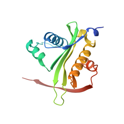
    Aminoglycoside N-acetyltransferases (AACs) confer resistance against the clinical use of aminoglycoside antibiotics. The origin of AACs can be traced to environmental microbial species representing a vast reservoir for new and emerging resistance enzymes, which are currently undercharacterized. Here, we performed detailed structural characterization and functional analyses of four metagenomic AAC (meta-AACs) enzymes recently identified in a survey of agricultural and grassland soil microbiomes ( Forsberg et al. Nature 2014 , 509 , 612 ). These enzymes are new members of the Gcn5-Related-N-Acetyltransferase superfamily and confer resistance to the aminoglycosides gentamicin C, sisomicin, and tobramycin. Moreover, the meta-AAC0020 enzyme demonstrated activity comparable with an AAC(3)-I enzyme that serves as a model AAC enzyme identified in a clinical bacterial isolate. The crystal structure of meta-AAC0020 in complex with sisomicin confirmed an unexpected AAC(6') regiospecificity of this enzyme and revealed a drug binding mechanism distinct from previously characterized AAC(6') enzymes. Together, our data highlights the presence of highly active antibiotic-modifying enzymes in the environmental microbiome and reveals unexpected diversity in substrate specificity. These observations of additional AAC enzymes must be considered in the search for novel aminoglycosides less prone to resistance.


  • Organizational Affiliation

    Department of Chemical Engineering and Applied Chemistry, University of Toronto , 200 College Street, Room 333, Toronto, Ontario M5S 3E5, Canada.


Macromolecules
Find similar proteins by:  (by identity cutoff)  |  3D Structure
Entity ID: 1
MoleculeChains Sequence LengthOrganismDetailsImage
aminoglycoside acetyltransferase meta-AAC0020
A, B, C, D
157uncultured bacteriumMutation(s): 1 
UniProt
Find proteins for A0A059WZ16 (uncultured bacterium)
Explore A0A059WZ16 
Go to UniProtKB:  A0A059WZ16
Entity Groups  
Sequence Clusters30% Identity50% Identity70% Identity90% Identity95% Identity100% Identity
UniProt GroupA0A059WZ16
Sequence Annotations
Expand
  • Reference Sequence
Small Molecules
Ligands 4 Unique
IDChains Name / Formula / InChI Key2D Diagram3D Interactions
SIS
Query on SIS

Download Ideal Coordinates CCD File 
E [auth A],
H [auth A],
M [auth C],
Q [auth D]
(1S,2S,3R,4S,6R)-4,6-diamino-3-{[(2S,3R)-3-amino-6-(aminomethyl)-3,4-dihydro-2H-pyran-2-yl]oxy}-2-hydroxycyclohexyl 3-deoxy-4-C-methyl-3-(methylamino)-beta-L-arabinopyranoside
C19 H37 N5 O7
URWAJWIAIPFPJE-YFMIWBNJSA-N
ACT
Query on ACT

Download Ideal Coordinates CCD File 
G [auth A],
L [auth B],
P [auth C],
R [auth D]
ACETATE ION
C2 H3 O2
QTBSBXVTEAMEQO-UHFFFAOYSA-M
CA
Query on CA

Download Ideal Coordinates CCD File 
I [auth A],
J [auth A],
K [auth B],
N [auth C],
O [auth C]
CALCIUM ION
Ca
BHPQYMZQTOCNFJ-UHFFFAOYSA-N
MG
Query on MG

Download Ideal Coordinates CCD File 
F [auth A],
S [auth D],
T [auth D]
MAGNESIUM ION
Mg
JLVVSXFLKOJNIY-UHFFFAOYSA-N
Experimental Data & Validation

Experimental Data

  • Method: X-RAY DIFFRACTION
  • Resolution: 1.52 Å
  • R-Value Free: 0.200 
  • R-Value Work: 0.166 
  • R-Value Observed: 0.167 
  • Space Group: P 1
  • Diffraction Data: https://doi.org/10.18430/M35U08
Unit Cell:
Length ( Å )Angle ( ˚ )
a = 46.295α = 71.76
b = 53.035β = 75.61
c = 78.676γ = 88.69
Software Package:
Software NamePurpose
PHENIXrefinement
XDSdata reduction
Aimlessdata scaling
PHENIXphasing
Cootmodel building

Structure Validation

View Full Validation Report


Ligand Structure Quality Assessment 


Entry History & Funding Information

Deposition Data


Funding OrganizationLocationGrant Number
National Institutes of Health/National Institute Of Allergy and Infectious Diseases (NIH/NIAID)United StatesHHSN272201200026C

Revision History  (Full details and data files)

  • Version 1.0: 2017-02-08
    Type: Initial release
  • Version 1.1: 2017-09-13
    Changes: Author supporting evidence
  • Version 1.2: 2019-04-03
    Changes: Data collection, Database references, Structure summary
  • Version 1.3: 2019-12-11
    Changes: Author supporting evidence
  • Version 1.4: 2023-10-04
    Changes: Data collection, Database references, Derived calculations, Refinement description