Structural insights into the cold adaptation of the photosynthetic pigment-protein C-phycocyanin from an Arctic cyanobacterium
Su, H.N., Wang, Q.M., Li, C.Y., Li, K., Luo, W., Chen, B., Zhang, X.Y., Qin, Q.L., Zhou, B.C., Chen, X.L., Zhang, Y.Z., Xie, B.B.(2017) Biochim Biophys Acta 1858: 325-335
- PubMed: 28188780 Search on PubMed
- DOI: https://doi.org/10.1016/j.bbabio.2017.02.004
- Primary Citation Related Structures:
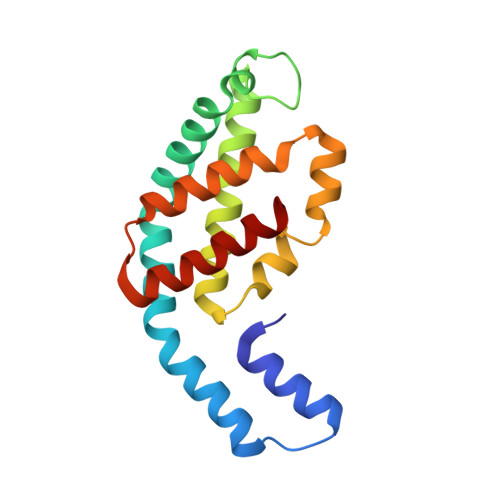
5TOU - PubMed Abstract:
The cold adaptation mechanism of phycobiliproteins, the major photosynthetic pigment-proteins in cyanobacteria and red algae, has rarely been studied. Here we reported the biochemical, structural, and molecular dynamics simulation study of the C-phycocyanin from Arctic cyanobacterial strain Pseudanabaena sp. LW0831. We characterized the phycobilisome components of LW0831 and obtained their gene sequences. Compared to the mesophilic counterpart from Arthrospira platensis (Ar-C-PC), LW0831 C-phycocyanin (Ps-C-PC) has a decreased thermostability (∆T m of -16°C), one of the typical features of cold-adapted enzymes. To uncover its structural basis, we resolved the crystal structure of Ps-C-PC 1 at 2.04Å. Consistent with the decrease in thermostability, comparative structural analyses revealed decreased intra-trimer and inter-trimer interactions in Ps-C-PC 1, compared to Ar-C-PC. However, comparative molecular dynamics simulations indicated that Ps-C-PC 1 shows similar flexibilities to Ar-C-PC for both the (αβ) 3 trimer and (αβ) 6 hexamer. Therefore, the optimization mode is clearly different from cold-adapted enzymes, which usually have increased flexibilities. Detailed analyses demonstrated different optimization modes for the α and β subunits and it was revealed that hydrophobic interactions are key to this difference, though salt bridges, hydrogen bonds, and surface hydrophobicity are also involved. This study is the first report of the structure of cold-adapted phycobiliproteins and provides insights into the cold-adaptation strategies of non-enzyme proteins.
- State Key Laboratory of Microbial Technology, Institute of Marine Science and Technology, Marine Biotechnology Research Center, Shandong University, Jinan 250100, China.
Organizational Affiliation: