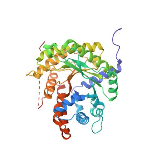
5TKL

Crystal structure of FBP aldolase from Toxoplasma gondii, condensation intermediate


Experimental Data Snapshot

  • Method: X-RAY DIFFRACTION
  • Resolution: 1.75 Å
  • R-Value Free: 0.177 
  • R-Value Work: 0.148 
  • R-Value Observed: 0.150 

wwPDB Validation   3D Report Full Report


Ligand Structure Quality Assessment 


This is version 1.3 of the entry. See complete history


Literature

Isomer activation controls stereospecificity of class I fructose-1,6-bisphosphate aldolases.

Heron, P.W.Sygusch, J.

(2017) J Biol Chem 292: 19849-19860

  • DOI: https://doi.org/10.1074/jbc.M117.811034
  • Primary Citation of Related Structures:  
    5TJS, 5TK3, 5TKC, 5TKL, 5TKN, 5TKP

  • PubMed Abstract: 

    Fructose-1,6-bisphosphate (FBP) aldolase, a glycolytic enzyme, catalyzes the reversible and stereospecific aldol addition of dihydroxyacetone phosphate (DHAP) and d-glyceraldehyde 3-phosphate (d-G3P) by an unresolved mechanism. To afford insight into the molecular determinants of FBP aldolase stereospecificity during aldol addition, a key ternary complex formed by DHAP and d-G3P, comprising 2% of the equilibrium population at physiological pH, was cryotrapped in the active site of Toxoplasma gondii aldolase crystals to high resolution. The growth of T. gondii aldolase crystals in acidic conditions enabled trapping of the ternary complex as a dominant population. The obligate 3( S )-4( R ) stereochemistry at the nascent C3-C4 bond of FBP requires a si -face attack by the covalent DHAP nucleophile on the d-G3P aldehyde si -face in the active site. The cis -isomer of the d-G3P aldehyde, representing the dominant population trapped in the ternary complex, would lead to re -face attack on the aldehyde and yield tagatose 1,6-bisphosphate, a competitive inhibitor of the enzyme. We propose that unhindered rotational isomerization by the d-G3P aldehyde moiety in the ternary complex generates the active trans -isomer competent for carbonyl bond activation by active-site residues, thereby enabling si -face attack by the DHAP enamine. C-C bond formation by the cis -isomer is suppressed by hydrogen bonding of the cis -aldehyde carbonyl with the DHAP enamine phosphate dianion through a tetrahedrally coordinated water molecule. The active site geometry further suppresses C-C bond formation with the l-G3P enantiomer of d-G3P. Understanding C-C formation is of fundamental importance in biological reactions and has considerable relevance to biosynthetic reactions in organic chemistry.


  • Organizational Affiliation

    From the Department of Biochemistry and Molecular Medicine, Université de Montréal, Montréal, Québec H3C 3J7, Canada.


Macromolecules
Find similar proteins by:  (by identity cutoff)  |  3D Structure
Entity ID: 1
MoleculeChains Sequence LengthOrganismDetailsImage
Fructose-bisphosphate aldolase363Toxoplasma gondiiMutation(s): 0 
Gene Names: ald-1
EC: 4.1.2.13
UniProt
Find proteins for Q8I8I2 (Toxoplasma gondii)
Explore Q8I8I2 
Go to UniProtKB:  Q8I8I2
Entity Groups  
Sequence Clusters30% Identity50% Identity70% Identity90% Identity95% Identity100% Identity
UniProt GroupQ8I8I2
Sequence Annotations
Expand
  • Reference Sequence
Small Molecules
Ligands 3 Unique
IDChains Name / Formula / InChI Key2D Diagram3D Interactions
P6F
Query on P6F

Download Ideal Coordinates CCD File 
B [auth A]1,6-di-O-phosphono-D-fructose
C6 H14 O12 P2
XPYBSIWDXQFNMH-UYFOZJQFSA-N
13P
Query on 13P

Download Ideal Coordinates CCD File 
C [auth A]1,3-DIHYDROXYACETONEPHOSPHATE
C3 H7 O6 P
GNGACRATGGDKBX-UHFFFAOYSA-N
G3H
Query on G3H

Download Ideal Coordinates CCD File 
D [auth A]GLYCERALDEHYDE-3-PHOSPHATE
C3 H7 O6 P
LXJXRIRHZLFYRP-VKHMYHEASA-N
Experimental Data & Validation

Experimental Data

  • Method: X-RAY DIFFRACTION
  • Resolution: 1.75 Å
  • R-Value Free: 0.177 
  • R-Value Work: 0.148 
  • R-Value Observed: 0.150 
  • Space Group: P 42 21 2
Unit Cell:
Length ( Å )Angle ( ˚ )
a = 109.621α = 90
b = 109.621β = 90
c = 54.958γ = 90
Software Package:
Software NamePurpose
PHENIXrefinement
CBASSdata collection
DENZOdata reduction
SCALEPACKdata scaling
PHENIXphasing
PDB_EXTRACTdata extraction

Structure Validation

View Full Validation Report


Ligand Structure Quality Assessment 


Entry History & Funding Information

Deposition Data


Funding OrganizationLocationGrant Number
Natural Sciences and Engineering Research Council (NSERC, Canada)Canada--

Revision History  (Full details and data files)

  • Version 1.0: 2017-10-04
    Type: Initial release
  • Version 1.1: 2017-10-18
    Changes: Database references
  • Version 1.2: 2017-12-13
    Changes: Database references
  • Version 1.3: 2020-01-08
    Changes: Author supporting evidence