5SXG

Crystal Structure of the Cancer Genomic DNA Mutator APOBEC3B


Experimental Data Snapshot

  • Method: X-RAY DIFFRACTION
  • Resolution: 1.93 Å
  • R-Value Free: 0.210 
  • R-Value Work: 0.177 
  • R-Value Observed: 0.178 

wwPDB Validation   3D Report Full Report


This is version 1.2 of the entry. See complete history


Literature

Conformational Switch Regulates the DNA Cytosine Deaminase Activity of Human APOBEC3B.

Shi, K.Demir, O.Carpenter, M.A.Wagner, J.Kurahashi, K.Harris, R.S.Amaro, R.E.Aihara, H.

(2017) Sci Rep 7: 17415-17415

  • DOI: https://doi.org/10.1038/s41598-017-17694-3
  • Primary Citation of Related Structures:  
    5SXG, 5SXH

  • PubMed Abstract: 

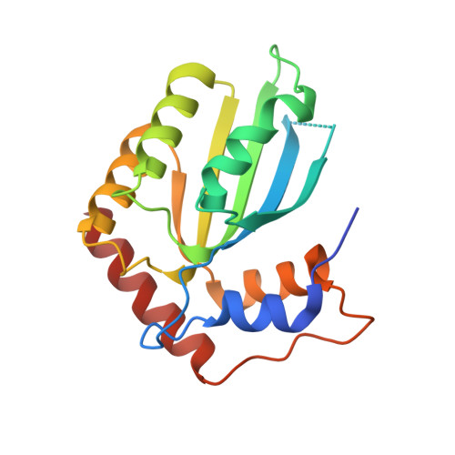
    The APOBEC3B (A3B) single-stranded DNA (ssDNA) cytosine deaminase has important roles in innate immunity but is also a major endogenous source of mutations in cancer. Previous structural studies showed that the C-terminal catalytic domain of human A3B has a tightly closed active site, and rearrangement of the surrounding loops is required for binding to substrate ssDNA. Here we report structures of the A3B catalytic domain in a new crystal form that show alternative, yet still closed, conformations of active site loops. All-atom molecular dynamics simulations support the dynamic behavior of active site loops and recapitulate the distinct modes of interactions that maintain a closed active site. Replacing segments of A3B loop 1 to mimic the more potent cytoplasmic deaminase APOBEC3A leads to elevated ssDNA deaminase activity, likely by facilitating opening of the active site. These data collectively suggest that conformational equilibrium of the A3B active site loops, skewed toward being closed, controls enzymatic activity by regulating binding to ssDNA substrates.


  • Organizational Affiliation

    Department of Biochemistry, Molecular Biology and Biophysics, University of Minnesota, Minneapolis, Minnesota, 55455, USA.


Macromolecules
Find similar proteins by:  (by identity cutoff)  |  3D Structure
Entity ID: 1
MoleculeChains Sequence LengthOrganismDetailsImage
DNA dC->dU-editing enzyme APOBEC-3B
A, B
185Homo sapiensMutation(s): 5 
Gene Names: APOBEC3B
EC: 3.5.4
UniProt & NIH Common Fund Data Resources
Find proteins for Q9UH17 (Homo sapiens)
Explore Q9UH17 
Go to UniProtKB:  Q9UH17
PHAROS:  Q9UH17
GTEx:  ENSG00000179750 
Entity Groups  
Sequence Clusters30% Identity50% Identity70% Identity90% Identity95% Identity100% Identity
UniProt GroupQ9UH17
Sequence Annotations
Expand
  • Reference Sequence
Small Molecules
Ligands 3 Unique
IDChains Name / Formula / InChI Key2D Diagram3D Interactions
PDO
Query on PDO

Download Ideal Coordinates CCD File 
D [auth A]
F [auth A]
H [auth B]
I [auth B]
J [auth B]
D [auth A],
F [auth A],
H [auth B],
I [auth B],
J [auth B],
K [auth B]
1,3-PROPANDIOL
C3 H8 O2
YPFDHNVEDLHUCE-UHFFFAOYSA-N
IMD
Query on IMD

Download Ideal Coordinates CCD File 
E [auth A]IMIDAZOLE
C3 H5 N2
RAXXELZNTBOGNW-UHFFFAOYSA-O
ZN
Query on ZN

Download Ideal Coordinates CCD File 
C [auth A],
G [auth B]
ZINC ION
Zn
PTFCDOFLOPIGGS-UHFFFAOYSA-N
Experimental Data & Validation

Experimental Data

  • Method: X-RAY DIFFRACTION
  • Resolution: 1.93 Å
  • R-Value Free: 0.210 
  • R-Value Work: 0.177 
  • R-Value Observed: 0.178 
  • Space Group: P 1 21 1
Unit Cell:
Length ( Å )Angle ( ˚ )
a = 44.39α = 90
b = 50.8β = 102.86
c = 76.89γ = 90
Software Package:
Software NamePurpose
PHENIXrefinement
XDSdata reduction
Aimlessdata scaling
PHASERphasing

Structure Validation

View Full Validation Report


Entry History & Funding Information

Deposition Data


Funding OrganizationLocationGrant Number
National Institutes of Health/National Institute of General Medical Sciences (NIH/NIGMS)United StatesGM095558 and GM109770

Revision History  (Full details and data files)

  • Version 1.0: 2017-12-27
    Type: Initial release
  • Version 1.1: 2019-12-25
    Changes: Author supporting evidence
  • Version 1.2: 2023-10-04
    Changes: Data collection, Database references, Derived calculations, Experimental preparation, Refinement description