5OY3

The structural basis of the histone demethylase KDM6B histone 3 lysine 27 specificity


Experimental Data Snapshot

  • Method: X-RAY DIFFRACTION
  • Resolution: 2.14 Å
  • R-Value Free: 0.221 
  • R-Value Work: 0.165 
  • R-Value Observed: 0.169 

wwPDB Validation   3D Report Full Report


This is version 1.2 of the entry. See complete history


Literature

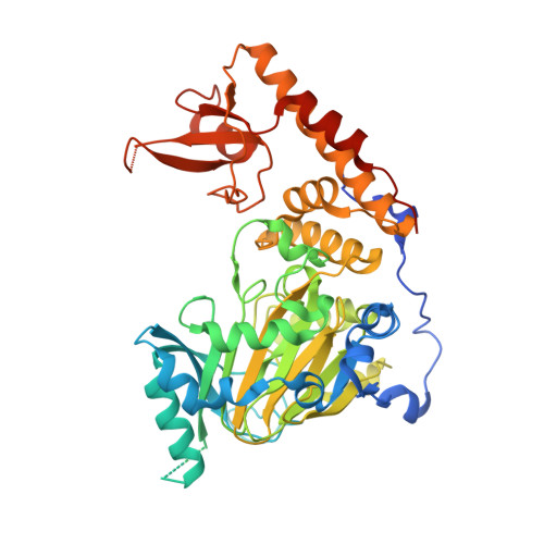
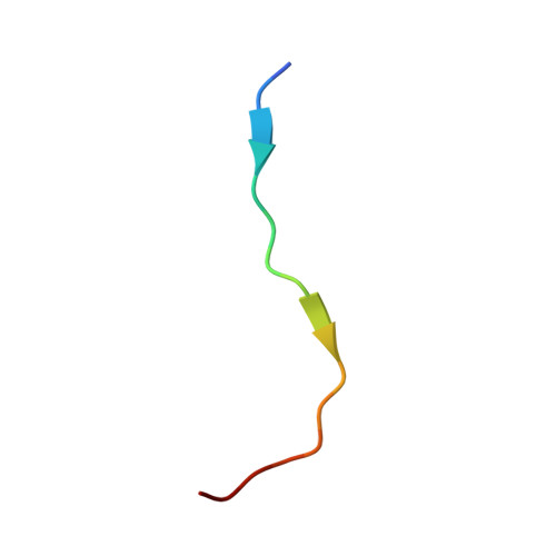
Structural Basis of Histone Demethylase KDM6B Histone 3 Lysine 27 Specificity.

Jones, S.E.Olsen, L.Gajhede, M.

(2018) Biochemistry 57: 585-592

  • DOI: https://doi.org/10.1021/acs.biochem.7b01152
  • Primary Citation of Related Structures:  
    5OY3, 6F6D

  • PubMed Abstract: 

    KDM subfamily 6 enzymes KDM6A and KDM6B specifically catalyze demethylation of di- and trimethylated lysine on histone 3 lysine 27 (H3K27me3/2) and play an important role in repression of developmental genes. Despite identical amino acid sequence in the immediate surroundings of H3K9me3/2 (ARKS), the enzymes do not catalyze demethylation of this general marker of repression. To address this question for KDM6B, we used computational methods to identify H3(17-33)-derived peptides with improved binding affinity that would allow co-crystallization with the catalytic core of human KDM6B (ccKDM6B). A total of five peptides were identified, and their IC 50 values were determined in a matrix-assisted laser desorption ionization time-of-flight-based assay. Despite none of the peptides showing affinity significantly higher than that of the H3(17-33) peptide, it was possible to co-crystallize ccKDM6B with a H3(17-33)A21M peptide. This structure reveals the interactions between the KDM6B zinc binding domain and the H3(17-23) region. Although KDM6A and KDM6B differ in primary sequence, particularly in the H3L20 binding pocket of the zinc binding domains, where two histidines in KDM6A have been replaced by a glutamate and a tyrosine, they bind H3(17-23) in a very similar fashion. This structure shows that KDM6B, in analogy with KDM6A, also uses the zinc binding domain to achieve H3K27me3/me2 specificity. The histidine to glutamine substitution at amino acid position 1564 in the KDM6B zinc binding domain can further explain why KDM6B binds substrates with an affinity higher than that of KDM6A.


  • Organizational Affiliation

    Biostructural Research, Department of Drug Design and Pharmacology, Faculty of Health and Medical Sciences, University of Copenhagen , Jagtvej 162, 2100 Copenhagen, Denmark.


Macromolecules
Find similar proteins by:  (by identity cutoff)  |  3D Structure
Entity ID: 1
MoleculeChains Sequence LengthOrganismDetailsImage
Lysine-specific demethylase 6B509Homo sapiensMutation(s): 0 
Gene Names: KDM6BJMJD3KIAA0346
EC: 1.14.11
UniProt & NIH Common Fund Data Resources
Find proteins for O15054 (Homo sapiens)
Explore O15054 
Go to UniProtKB:  O15054
PHAROS:  O15054
GTEx:  ENSG00000132510 
Entity Groups  
Sequence Clusters30% Identity50% Identity70% Identity90% Identity95% Identity100% Identity
UniProt GroupO15054
Sequence Annotations
Expand
  • Reference Sequence

Find similar proteins by:  Sequence   |   3D Structure  

Entity ID: 2
MoleculeChains Sequence LengthOrganismDetailsImage
Histone 3 peptide H3(17-33)A21M17Homo sapiensMutation(s): 0 
UniProt & NIH Common Fund Data Resources
Find proteins for P68431 (Homo sapiens)
Explore P68431 
Go to UniProtKB:  P68431
PHAROS:  P68431
Entity Groups  
Sequence Clusters30% Identity50% Identity70% Identity90% Identity95% Identity100% Identity
UniProt GroupP68431
Sequence Annotations
Expand
  • Reference Sequence
Small Molecules
Ligands 5 Unique
IDChains Name / Formula / InChI Key2D Diagram3D Interactions
AKG
Query on AKG

Download Ideal Coordinates CCD File 
E [auth A]2-OXOGLUTARIC ACID
C5 H6 O5
KPGXRSRHYNQIFN-UHFFFAOYSA-N
TBU
Query on TBU

Download Ideal Coordinates CCD File 
N [auth A]TERTIARY-BUTYL ALCOHOL
C4 H10 O
DKGAVHZHDRPRBM-UHFFFAOYSA-N
ZN
Query on ZN

Download Ideal Coordinates CCD File 
D [auth A]ZINC ION
Zn
PTFCDOFLOPIGGS-UHFFFAOYSA-N
EDO
Query on EDO

Download Ideal Coordinates CCD File 
F [auth A]
G [auth A]
H [auth A]
I [auth A]
J [auth A]
F [auth A],
G [auth A],
H [auth A],
I [auth A],
J [auth A],
K [auth A],
L [auth A],
M [auth A]
1,2-ETHANEDIOL
C2 H6 O2
LYCAIKOWRPUZTN-UHFFFAOYSA-N
FE
Query on FE

Download Ideal Coordinates CCD File 
C [auth A]FE (III) ION
Fe
VTLYFUHAOXGGBS-UHFFFAOYSA-N
Experimental Data & Validation

Experimental Data

  • Method: X-RAY DIFFRACTION
  • Resolution: 2.14 Å
  • R-Value Free: 0.221 
  • R-Value Work: 0.165 
  • R-Value Observed: 0.169 
  • Space Group: P 41 21 2
Unit Cell:
Length ( Å )Angle ( ˚ )
a = 68.908α = 90
b = 68.908β = 90
c = 228.476γ = 90
Software Package:
Software NamePurpose
PHENIXrefinement
XDSdata reduction
Aimlessdata scaling
PHASERphasing

Structure Validation

View Full Validation Report


Entry History & Funding Information

Deposition Data


Funding OrganizationLocationGrant Number
Danish Council for Independent ResearchDenmark--

Revision History  (Full details and data files)

  • Version 1.0: 2017-09-20
    Type: Initial release
  • Version 1.1: 2019-04-03
    Changes: Author supporting evidence, Data collection, Database references
  • Version 1.2: 2024-01-17
    Changes: Data collection, Database references, Refinement description