Structure and function of the type III pullulan hydrolase from Thermococcus kodakarensis.
Guo, J., Coker, A.R., Wood, S.P., Cooper, J.B., Keegan, R.M., Ahmad, N., Muhammad, M.A., Rashid, N., Akhtar, M.(2018) Acta Crystallogr D Struct Biol 74: 305-314
- PubMed: 29652257 Search on PubMed
- DOI: https://doi.org/10.1107/S2059798318001754
- Primary Citation Related Structures:
5OT1 - PubMed Abstract:
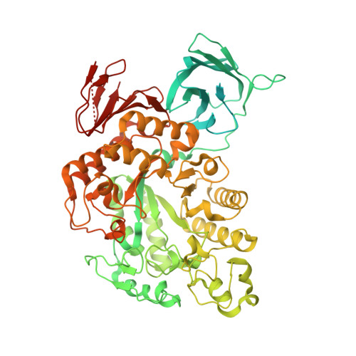
Pullulan-hydrolysing enzymes, more commonly known as debranching enzymes for starch and other polysaccharides, are of great interest and have been widely used in the starch-saccharification industry. Type III pullulan hydrolase from Thermococcus kodakarensis (TK-PUL) possesses both pullulanase and α-amylase activities. Until now, only two enzymes in this class, which are capable of hydrolysing both α-1,4- and α-1,6-glycosidic bonds in pullulan to produce a mixture of maltose, panose and maltotriose, have been described. TK-PUL shows highest activity in the temperature range 95-100°C and has a pH optimum in the range 3.5-4.2. Its unique ability to hydrolyse maltotriose into maltose and glucose has not been reported for other homologous enzymes. The crystal structure of TK-PUL has been determined at a resolution of 2.8 Å and represents the first analysis of a type III pullulan hydrolyse. The structure reveals that the last part of the N-terminal domain and the C-terminal domain are significantly different from homologous structures. In addition, the loop regions at the active-site end of the central catalytic domain are quite different. The enzyme has a well defined calcium-binding site and possesses a rare vicinal disulfide bridge. The thermostability of TK-PUL and its homologues may be attributable to several factors, including the increased content of salt bridges, helical segments, Pro, Arg and Tyr residues and the decreased content of serine.
- Division of Medicine, University College London, Gower Street, London WC1E 6BT, England.
Organizational Affiliation: