5OLL | pdb_00005oll

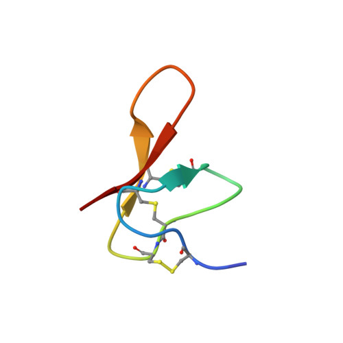
Crystal structure of gurmarin, a sweet taste suppressing polypeptide


Experimental Data Snapshot

  • Method: X-RAY DIFFRACTION
  • Resolution: 1.45 Å
  • R-Value Free: 
    0.191 (Depositor), 0.217 (DCC) 
  • R-Value Work: 
    0.190 (Depositor), 0.192 (DCC) 
  • R-Value Observed: 
    0.190 (Depositor) 

wwPDB Validation 3D Report Full Report

Validation slider image for 5OLL

This is version 1.2 of the entry. See complete history. 

Literature

The Crystal Structure of Gurmarin, a Sweet Taste-Suppressing Protein: Identification of the Amino Acid Residues Essential for Inhibition.

Sigoillot, M., Brockhoff, A., Neiers, F., Poirier, N., Belloir, C., Legrand, P., Charron, C., Roblin, P., Meyerhof, W., Briand, L.

(2018) Chem Senses 43: 635-643

  • DOI: https://doi.org/10.1093/chemse/bjy054
  • Primary Citation Related Structures: 
    5OLL

  • PubMed Abstract: 

    Gurmarin is a highly specific sweet taste-suppressing protein in rodents that is isolated from the Indian plant Gymnema sylvestre. Gurmarin consists of 35 amino acid residues containing 3 intramolecular disulfide bridges that form a cystine knot. Here, we report the crystal structure of gurmarin at a 1.45 Å resolution and compare it with previously reported nuclear magnetic resonance solution structures. The atomic structure at this resolution allowed us to identify a very flexible region consisting of hydrophobic residues. Some of these amino acid residues had been identified as a putative binding site for the rat sweet taste receptor in a previous study. By combining alanine-scanning mutagenesis of the gurmarin molecule and a functional cell-based receptor assay, we confirmed that some single point mutations in these positions drastically affect sweet taste receptor inhibition by gurmarin.


  • Organizational Affiliation: 
    • INRA, CNRS, Centre des Sciences du Goût et de l'Alimentation, Université de Bourgogne-Franche Comté, Dijon, France.

Macromolecule Content 

  • Total Structure Weight: 4.42 kDa 
  • Atom Count: 367 
  • Modeled Residue Count: 35 
  • Deposited Residue Count: 35 
  • Unique protein chains: 1

Macromolecules

Find similar proteins by:|  3D Structure
Entity ID: 1
MoleculeChains  Sequence LengthOrganismDetailsImage
Gurmarin35Gymnema sylvestreMutation(s): 0 
UniProt
Find proteins for P25810 (Gymnema sylvestre)
Explore P25810 
Go to UniProtKB:  P25810
Entity Groups
Sequence Clusters30% Identity50% Identity70% Identity90% Identity95% Identity100% Identity
UniProt GroupP25810
Sequence Annotations
Expand
Reference Sequence

Experimental Data & Validation

Experimental Data

  • Method: X-RAY DIFFRACTION
  • Resolution: 1.45 Å
  • R-Value Free:  0.191 (Depositor), 0.217 (DCC) 
  • R-Value Work:  0.190 (Depositor), 0.192 (DCC) 
  • R-Value Observed: 0.190 (Depositor) 
Space Group: H 3
Unit Cell:
Length ( Å )Angle ( ˚ )
a = 53.65α = 90
b = 53.65β = 90
c = 38.11γ = 120
Software Package:
Software NamePurpose
BUSTERrefinement
XDSdata reduction
XDSdata scaling
SHELXDEphasing

Structure Validation

View Full Validation Report


Entry History 

Deposition Data

Revision History  (Full details and data files)

  • Version 1.0: 2018-08-08
    Type: Initial release
  • Version 1.1: 2019-02-20
    Changes: Data collection, Database references
  • Version 1.2: 2024-11-20
    Changes: Data collection, Database references, Derived calculations, Structure summary