5OHY

A GH31 family sulfoquinovosidase in complex with aza-sugar inhibitor IFGSQ


Experimental Data Snapshot

  • Method: X-RAY DIFFRACTION
  • Resolution: 1.77 Å
  • R-Value Free: 0.195 
  • R-Value Work: 0.177 
  • R-Value Observed: 0.178 

wwPDB Validation   3D Report Full Report


Ligand Structure Quality Assessment 


This is version 1.2 of the entry. See complete history


Literature

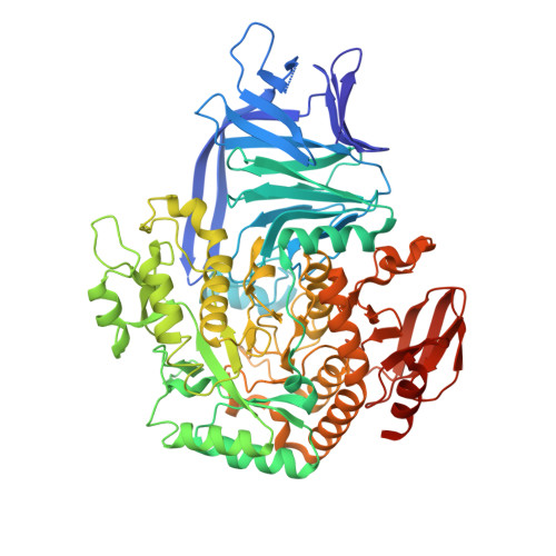
Structural and Biochemical Insights into the Function and Evolution of Sulfoquinovosidases.

Abayakoon, P.Jin, Y.Lingford, J.P.Petricevic, M.John, A.Ryan, E.Wai-Ying Mui, J.Pires, D.E.V.Ascher, D.B.Davies, G.J.Goddard-Borger, E.D.Williams, S.J.

(2018) ACS Cent Sci 4: 1266-1273

  • DOI: https://doi.org/10.1021/acscentsci.8b00453
  • Primary Citation of Related Structures:  
    5OHS, 5OHT, 5OHY

  • PubMed Abstract: 

    An estimated 10 billion tonnes of sulfoquinovose (SQ) are produced and degraded each year. Prokaryotic sulfoglycolytic pathways catabolize sulfoquinovose (SQ) liberated from plant sulfolipid, or its delipidated form α-d-sulfoquinovosyl glycerol (SQGro), through the action of a sulfoquinovosidase (SQase), but little is known about the capacity of SQ glycosides to support growth. Structural studies of the first reported SQase ( Escherichia coli YihQ) have identified three conserved residues that are essential for substrate recognition, but crossover mutations exploring active-site residues of predicted SQases from other organisms have yielded inactive mutants casting doubt on bioinformatic functional assignment. Here, we show that SQGro can support the growth of E. coli on par with d-glucose, and that the E. coli SQase prefers the naturally occurring diastereomer of SQGro. A predicted, but divergent, SQase from Agrobacterium tumefaciens proved to have highly specific activity toward SQ glycosides, and structural, mutagenic, and bioinformatic analyses revealed the molecular coevolution of catalytically important amino acid pairs directly involved in substrate recognition, as well as structurally important pairs distal to the active site. Understanding the defining features of SQases empowers bioinformatic approaches for mapping sulfur metabolism in diverse microbial communities and sheds light on this poorly understood arm of the biosulfur cycle.


  • Organizational Affiliation

    School of Chemistry and Bio21 Molecular Science and Biotechnology Institute, University of Melbourne, Parkville, Victoria 3010, Australia.


Macromolecules
Find similar proteins by:  (by identity cutoff)  |  3D Structure
Entity ID: 1
MoleculeChains Sequence LengthOrganismDetailsImage
Alpha-glucosidase yihQ
A, B, C, D
672Agrobacterium tumefaciensMutation(s): 2 
Gene Names: SY94_3281
EC: 3.2.1.20
UniProt
Find proteins for A9CEZ0 (Agrobacterium fabrum (strain C58 / ATCC 33970))
Explore A9CEZ0 
Go to UniProtKB:  A9CEZ0
Entity Groups  
Sequence Clusters30% Identity50% Identity70% Identity90% Identity95% Identity100% Identity
UniProt GroupA9CEZ0
Sequence Annotations
Expand
  • Reference Sequence
Small Molecules
Ligands 5 Unique
IDChains Name / Formula / InChI Key2D Diagram3D Interactions
9VH
Query on 9VH

Download Ideal Coordinates CCD File 
DA [auth D],
H [auth A],
P [auth B],
X [auth C]
[(3~{S},4~{R},5~{R})-4,5-bis(oxidanyl)piperidin-3-yl]methanesulfonic acid
C6 H13 N O5 S
RCSXCFCCYYFFCY-HSUXUTPPSA-N
PO4
Query on PO4

Download Ideal Coordinates CCD File 
I [auth A],
Q [auth B],
Y [auth C]
PHOSPHATE ION
O4 P
NBIIXXVUZAFLBC-UHFFFAOYSA-K
EDO
Query on EDO

Download Ideal Coordinates CCD File 
AA [auth D]
BA [auth D]
CA [auth D]
F [auth A]
K [auth B]
AA [auth D],
BA [auth D],
CA [auth D],
F [auth A],
K [auth B],
L [auth B],
M [auth B],
S [auth C],
T [auth C],
U [auth C]
1,2-ETHANEDIOL
C2 H6 O2
LYCAIKOWRPUZTN-UHFFFAOYSA-N
SCN
Query on SCN

Download Ideal Coordinates CCD File 
G [auth A],
N [auth B],
O [auth B],
V [auth C],
W [auth C]
THIOCYANATE ION
C N S
ZMZDMBWJUHKJPS-UHFFFAOYSA-M
K
Query on K

Download Ideal Coordinates CCD File 
E [auth A],
J [auth B],
R [auth C],
Z [auth D]
POTASSIUM ION
K
NPYPAHLBTDXSSS-UHFFFAOYSA-N
Experimental Data & Validation

Experimental Data

  • Method: X-RAY DIFFRACTION
  • Resolution: 1.77 Å
  • R-Value Free: 0.195 
  • R-Value Work: 0.177 
  • R-Value Observed: 0.178 
  • Space Group: P 1 21 1
Unit Cell:
Length ( Å )Angle ( ˚ )
a = 98.29α = 90
b = 168.82β = 117.07
c = 101.39γ = 90
Software Package:
Software NamePurpose
REFMACrefinement
xia2data reduction
Aimlessdata scaling
MOLREPphasing

Structure Validation

View Full Validation Report


Ligand Structure Quality Assessment 


Entry History 

Deposition Data

Revision History  (Full details and data files)

  • Version 1.0: 2018-08-08
    Type: Initial release
  • Version 1.1: 2019-02-20
    Changes: Data collection, Database references, Structure summary
  • Version 1.2: 2024-05-01
    Changes: Data collection, Database references, Derived calculations, Refinement description