5OFU

Crystal structure of Leishmania major fructose-1,6-bisphosphatase in T-state.


Experimental Data Snapshot

  • Method: X-RAY DIFFRACTION
  • Resolution: 2.62 Å
  • R-Value Free: 0.216 
  • R-Value Work: 0.189 
  • R-Value Observed: 0.190 

wwPDB Validation   3D Report Full Report


Ligand Structure Quality Assessment 


This is version 1.3 of the entry. See complete history


Literature

Structures of Leishmania Fructose-1,6-Bisphosphatase Reveal Species-Specific Differences in the Mechanism of Allosteric Inhibition.

Yuan, M.Vasquez-Valdivieso, M.G.McNae, I.W.Michels, P.A.M.Fothergill-Gilmore, L.A.Walkinshaw, M.D.

(2017) J Mol Biol 429: 3075-3089

  • DOI: https://doi.org/10.1016/j.jmb.2017.08.010
  • Primary Citation of Related Structures:  
    5OEY, 5OEZ, 5OFU

  • PubMed Abstract: 

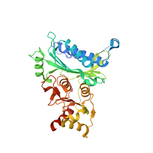
    The gluconeogenic enzyme fructose-1,6-bisphosphatase has been proposed as a potential drug target against Leishmania parasites that cause up to 20,000-30,000 deaths annually. A comparison of three crystal structures of Leishmania major fructose-1,6-bisphosphatase (LmFBPase) along with enzyme kinetic data show how AMP acts as an allosteric inhibitor and provides insight into its metal-dependent reaction mechanism. The crystal structure of the apoenzyme form of LmFBPase is a homotetramer in which the dimer of dimers adopts a planar conformation with disordered "dynamic loops". The structure of LmFBPase, complexed with manganese and its catalytic product phosphate, shows the dynamic loops locked into the active sites. A third crystal structure of LmFBPase complexed with its allosteric inhibitor AMP shows an inactive form of the tetramer, in which the dimer pairs are rotated by 18° relative to each other. The three structures suggest an allosteric mechanism in which AMP binding triggers a rearrangement of hydrogen bonds across the large and small interfaces. Retraction of the "effector loop" required for AMP binding releases the side chain of His23 from the dimer-dimer interface. This is coupled with a flip of the side chain of Arg48 which ties down the key catalytic dynamic loop in a disengaged conformation and also locks the tetramer in an inactive rotated T-state. The structure of the effector site of LmFBPase shows different structural features compared with human FBPases, thereby offering a potential and species-specific drug target.


  • Organizational Affiliation

    Centre for Translational and Chemical Biology, School of Biological Sciences, University of Edinburgh, Michael Swann Building, Max Born Crescent, Edinburgh EH9 3BF, UK.


Macromolecules
Find similar proteins by:  (by identity cutoff)  |  3D Structure
Entity ID: 1
MoleculeChains Sequence LengthOrganismDetailsImage
FBP protein
A, B, C, D
351Leishmania majorMutation(s): 0 
Gene Names: FBPLMJF_04_1160
EC: 3.1.3.11
UniProt
Find proteins for O97193 (Leishmania major)
Explore O97193 
Go to UniProtKB:  O97193
Entity Groups  
Sequence Clusters30% Identity50% Identity70% Identity90% Identity95% Identity100% Identity
UniProt GroupO97193
Sequence Annotations
Expand
  • Reference Sequence
Small Molecules
Ligands 3 Unique
IDChains Name / Formula / InChI Key2D Diagram3D Interactions
AMP
Query on AMP

Download Ideal Coordinates CCD File 
E [auth A],
H [auth B],
J [auth C],
M [auth D]
ADENOSINE MONOPHOSPHATE
C10 H14 N5 O7 P
UDMBCSSLTHHNCD-KQYNXXCUSA-N
F6P
Query on F6P

Download Ideal Coordinates CCD File 
F [auth A],
I [auth B],
K [auth C],
N [auth D]
6-O-phosphono-beta-D-fructofuranose
C6 H13 O9 P
BGWGXPAPYGQALX-ARQDHWQXSA-N
CL
Query on CL

Download Ideal Coordinates CCD File 
G [auth A],
L [auth C]
CHLORIDE ION
Cl
VEXZGXHMUGYJMC-UHFFFAOYSA-M
Experimental Data & Validation

Experimental Data

  • Method: X-RAY DIFFRACTION
  • Resolution: 2.62 Å
  • R-Value Free: 0.216 
  • R-Value Work: 0.189 
  • R-Value Observed: 0.190 
  • Space Group: P 21 21 21
Unit Cell:
Length ( Å )Angle ( ˚ )
a = 90.59α = 90
b = 109.25β = 90
c = 140.51γ = 90
Software Package:
Software NamePurpose
REFMACrefinement
xia2data reduction
Aimlessdata scaling
PHASERphasing

Structure Validation

View Full Validation Report


Ligand Structure Quality Assessment 


Entry History 

Deposition Data

Revision History  (Full details and data files)

  • Version 1.0: 2017-09-20
    Type: Initial release
  • Version 1.1: 2017-10-18
    Changes: Database references
  • Version 1.2: 2020-07-29
    Type: Remediation
    Reason: Carbohydrate remediation
    Changes: Data collection, Derived calculations, Structure summary
  • Version 1.3: 2024-01-17
    Changes: Data collection, Database references, Refinement description, Structure summary