5OD2 | pdb_00005od2

Crystal structure of ADP-dependent glucokinase from Methanocaldococcus jannaschii


Experimental Data Snapshot

  • Method: X-RAY DIFFRACTION
  • Resolution: 1.98 Å
  • R-Value Free: 
    0.224 (Depositor), 0.227 (DCC) 
  • R-Value Work: 
    0.186 (Depositor), 0.193 (DCC) 
  • R-Value Observed: 
    0.188 (Depositor) 

Starting Model: experimental
View more details

wwPDB Validation 3D Report Full Report

Validation slider image for 5OD2

Ligand Structure Quality Assessment 


This is version 1.3 of the entry. See complete history

Literature

Crystal structure of ADP-dependent glucokinase from Methanocaldococcus jannaschii in complex with 5-iodotubercidin reveals phosphoryl transfer mechanism.

Tokarz, P.Wisniewska, M.Kaminski, M.M.Dubin, G.Grudnik, P.

(2018) Protein Sci 27: 790-797

  • DOI: https://doi.org/10.1002/pro.3377
  • Primary Citation Related Structures: 
    5OD2

  • PubMed Abstract: 

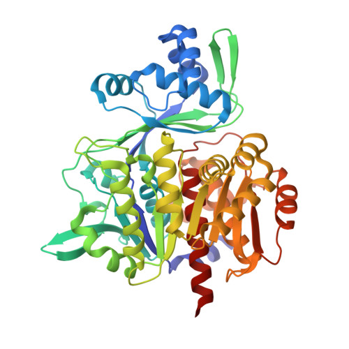
    ADP-dependent glucokinase (ADPGK) is an alternative novel glucose phosphorylating enzyme in a modified glycolysis pathway of hyperthermophilic Archaea. In contrast to classical ATP-dependent hexokinases, ADPGK utilizes ADP as a phosphoryl group donor. Here, we present a crystal structure of archaeal ADPGK from Methanocaldococcus jannaschii in complex with an inhibitor, 5-iodotubercidin, d-glucose, inorganic phosphate, and a magnesium ion. Detailed analysis of the architecture of the active site allowed for confirmation of the previously proposed phosphorylation mechanism and the crucial role of the invariant arginine residue (Arg197). The crystal structure shows how the phosphate ion, while mimicking a β-phosphate group, is positioned in the proximity of the glucose moiety by arginine and the magnesium ion, thus providing novel insights into the mechanism of catalysis. In addition, we demonstrate that 5-iodotubercidin inhibits human ADPGK-dependent T cell activation-induced reactive oxygen species (ROS) release and downstream gene expression, and as such it may serve as a model compound for further screening for hADPGK-specific inhibitors.


  • Organizational Affiliation
    • Faculty of Biochemistry, Biophysics and Biotechnology, Jagiellonian University in Krakow, ul. Gronostajowa 7, Krakow, 30-387, Poland.

Macromolecule Content 

  • Total Structure Weight: 161.09 kDa 
  • Atom Count: 11,882 
  • Modeled Residue Count: 1,370 
  • Deposited Residue Count: 1,374 
  • Unique protein chains: 1

Macromolecules

Find similar proteins by:|  3D Structure
Entity ID: 1
MoleculeChains  Sequence LengthOrganismDetailsImage
Bifunctional ADP-specific glucokinase/phosphofructokinase
A, B, C
458Methanocaldococcus jannaschii DSM 2661Mutation(s): 0 
Gene Names: pfkCglkAMJ1604
EC: 2.7.1.146 (PDB Primary Data), 2.7.1.147 (PDB Primary Data)
UniProt
Find proteins for Q58999 (Methanocaldococcus jannaschii (strain ATCC 43067 / DSM 2661 / JAL-1 / JCM 10045 / NBRC 100440))
Explore Q58999 
Go to UniProtKB:  Q58999
Entity Groups
Sequence Clusters30% Identity50% Identity70% Identity90% Identity95% Identity100% Identity
UniProt GroupQ58999
Sequence Annotations
Expand
Reference Sequence

Small Molecules

Ligands 4 Unique
IDChains Name / Formula / InChI Key2D Diagram3D Interactions
5ID

Query on 5ID


Download:Ideal Coordinates CCD File
E [auth A],
I [auth B],
M [auth C]
(2R,3R,4S,5R)-2-(4-AMINO-5-IODO-7H-PYRROLO[2,3-D]PYRIMIDIN-7-YL)-5-(HYDROXYMETHYL)TETRAHYDROFURAN-3,4-DIOL
C11 H13 I N4 O4
WHSIXKUPQCKWBY-IOSLPCCCSA-N
GLC

Query on GLC


Download:Ideal Coordinates CCD File
D [auth A],
H [auth B],
L [auth C]
alpha-D-glucopyranose
C6 H12 O6
WQZGKKKJIJFFOK-DVKNGEFBSA-N
PO4

Query on PO4


Download:Ideal Coordinates CCD File
G [auth A],
K [auth B],
O [auth C]
PHOSPHATE ION
O4 P
NBIIXXVUZAFLBC-UHFFFAOYSA-K
MG

Query on MG


Download:Ideal Coordinates CCD File
F [auth A],
J [auth B],
N [auth C]
MAGNESIUM ION
Mg
JLVVSXFLKOJNIY-UHFFFAOYSA-N

Experimental Data & Validation

Experimental Data

  • Method: X-RAY DIFFRACTION
  • Resolution: 1.98 Å
  • R-Value Free:  0.224 (Depositor), 0.227 (DCC) 
  • R-Value Work:  0.186 (Depositor), 0.193 (DCC) 
  • R-Value Observed: 0.188 (Depositor) 
Space Group: P 31
Unit Cell:
Length ( Å )Angle ( ˚ )
a = 154.565α = 90
b = 154.565β = 90
c = 50.483γ = 120
Software Package:
Software NamePurpose
REFMACrefinement
PDB_EXTRACTdata extraction
XDSdata reduction
XSCALEdata scaling
PHASERphasing

Structure Validation

View Full Validation Report


Ligand Structure Quality Assessment 


Entry History 

& Funding Information

Deposition Data


Funding OrganizationLocationGrant Number
National Science CentrePolandUMO-2015/19/D/NZ1/02009

Revision History  (Full details and data files)

  • Version 1.0: 2018-01-31
    Type: Initial release
  • Version 1.1: 2018-02-28
    Changes: Database references
  • Version 1.2: 2020-07-29
    Type: Remediation
    Reason: Carbohydrate remediation
    Changes: Data collection, Derived calculations, Structure summary
  • Version 1.3: 2024-01-17
    Changes: Data collection, Database references, Refinement description, Structure summary