Protein topology determines substrate-binding mechanism in homologous enzymes.
Herrera-Morande, A., Castro-Fernandez, V., Merino, F., Ramirez-Sarmiento, C.A., Fernandez, F.J., Vega, M.C., Guixe, V.(2018) Biochim Biophys Acta Gen Subj 1862: 2869-2878
- PubMed: 30251675 Search on PubMed
- DOI: https://doi.org/10.1016/j.bbagen.2018.09.007
- Primary Citation Related Structures:
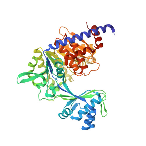
5O5X, 5O5Y, 5O5Z - PubMed Abstract:
During evolution, some homologs proteins appear with different connectivity between secondary structures (different topology) but conserving the tridimensional arrangement of them (same architecture). These events can produce two types of arrangements; circular permutation or non-cyclic permutations. The first one results in the N and C terminus transferring to a different position on a protein sequence while the second refers to a more complex arrangement of the structural elements. In ribokinase superfamily, two different topologies can be identified, which are related to each other as a non-cyclic permutation occurred during the evolution. Interestingly, this change in topology is correlated with the nucleotide specificity of its members. Thereby, the connectivity of the secondary elements allows us to distinguish an ATP-dependent and an ADP-dependent topology. Here we address the impact of introducing the topology of a homologous ATP-dependent kinase in an ADP-dependent kinase (Thermococcus litoralis glucokinase) in the structure, nucleotide specificity, and substrate binding order of the engineered enzyme. Structural evidence demonstrates that rewiring the topology of TlGK leads to an active and soluble enzyme without modifications on its three-dimensional architecture. The permuted enzyme (PerGK) retains the nucleotide preference of the parent TlGK enzyme but shows a change in the substrate binding order. Our results illustrate how the rearrangement of the protein folding topology during the evolution of the ribokinase superfamily enzymes may have dictated the substrate-binding order in homologous enzymes of this superfamily.
- Departamento de Biología, Facultad de Ciencias, Universidad de Chile, Santiago, Chile.
Organizational Affiliation: