Biochemical Function, Molecular Structure and Evolution of an Atypical Thioredoxin Reductase from Desulfovibrio vulgaris.
Valette, O., Tran, T.T.T., Cavazza, C., Caudeville, E., Brasseur, G., Dolla, A., Talla, E., Pieulle, L.(2017) Front Microbiol 8: 1855-1855
- PubMed: 29033913 Search on PubMedSearch on PubMed Central
- DOI: https://doi.org/10.3389/fmicb.2017.01855
- Primary Citation Related Structures:
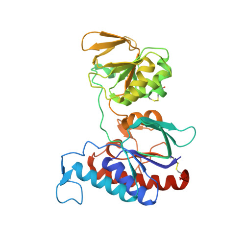
5NII - PubMed Abstract:
Thioredoxin reductase (TR) regulates the intracellular redox environment by reducing thioredoxin (Trx). In anaerobes, recent findings indicate that the Trx redox network is implicated in the global redox regulation of metabolism but also actively participates in protecting cells against O 2 . In the anaerobe Desulfovibrio vulgaris Hildenborough ( Dv H), there is an intriguing redundancy of the Trx system which includes a classical system using NADPH as electron source, a non-canonical system using NADH and an isolated TR (DvTRi). The functionality of DvTRi was questioned due to its lack of reactivity with DvTrxs. Structural analysis shows that DvTRi is a NAD(P)H-independent TR but its reducer needs still to be identified. Moreover, DvTRi reduced by an artificial electron source is able to reduce in turn DvTrx1 and complexation experiments demonstrate a direct interaction between DvTRi and DvTrx1. The deletion mutant tri exhibits a higher sensitivity to disulfide stress and the gene tri is upregulated by O 2 exposure. Having DvTRi in addition to DvTR1 as electron source for reducing DvTrx1 must be an asset to combat oxidative stress. Large-scale phylogenomics analyses show that TRi homologs are confined within the anaerobes. All TRi proteins displayed a conserved TQ/NGK motif instead of the HRRD motif, which is selective for the binding of the 2'-phosphate group of NADPH. The evolutionary history of TRs indicates that tr1 is the common gene ancestor in prokaryotes, affected by both gene duplications and horizontal gene events, therefore leading to the appearance of TRi through subfunctionalization over the evolutionary time.
- Aix-Marseille Univ, CNRS, LCB, Marseille, France.
Organizational Affiliation: