5MV2

Crystal structure of the E protein of the Japanese encephalitis live attenuated vaccine virus


Experimental Data Snapshot

  • Method: X-RAY DIFFRACTION
  • Resolution: 2.10 Å
  • R-Value Free: 0.223 
  • R-Value Work: 0.180 
  • R-Value Observed: 0.182 

Starting Model: experimental
View more details

wwPDB Validation   3D Report Full Report


This is version 1.2 of the entry. See complete history


Literature

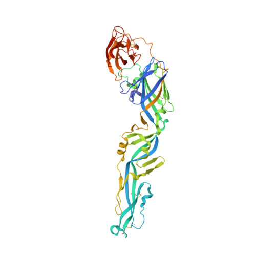
The structure differences of Japanese encephalitis virus SA14 and SA14-14-2 E proteins elucidate the virulence attenuation mechanism.

Liu, X.Zhao, X.Na, R.Li, L.Warkentin, E.Witt, J.Lu, X.Yu, Y.Wei, Y.Peng, G.Li, Y.Wang, J.

(2019) Protein Cell 10: 149-153


Macromolecules
Find similar proteins by:  (by identity cutoff)  |  3D Structure
Entity ID: 1
MoleculeChains Sequence LengthOrganismDetailsImage
E protein416Japanese encephalitis virus strain SA-14Mutation(s): 0 
Membrane Entity: Yes 
UniProt
Find proteins for P27395 (Japanese encephalitis virus (strain SA-14))
Explore P27395 
Go to UniProtKB:  P27395
Entity Groups  
Sequence Clusters30% Identity50% Identity70% Identity90% Identity95% Identity100% Identity
UniProt GroupP27395
Sequence Annotations
Expand
  • Reference Sequence
Experimental Data & Validation

Experimental Data

  • Method: X-RAY DIFFRACTION
  • Resolution: 2.10 Å
  • R-Value Free: 0.223 
  • R-Value Work: 0.180 
  • R-Value Observed: 0.182 
  • Space Group: C 1 2 1
Unit Cell:
Length ( Å )Angle ( ˚ )
a = 130.6α = 90
b = 55.9β = 132.1
c = 90.1γ = 90
Software Package:
Software NamePurpose
PHENIXrefinement
XDSdata reduction
XDSdata scaling
PHASERphasing

Structure Validation

View Full Validation Report


Entry History 

Deposition Data

Revision History  (Full details and data files)

  • Version 1.0: 2018-05-23
    Type: Initial release
  • Version 1.1: 2019-02-27
    Changes: Data collection, Database references
  • Version 1.2: 2024-01-17
    Changes: Data collection, Database references, Refinement description