5MFU | pdb_00005mfu

PA3825-EAL Mn-pGpG Structure


Experimental Data Snapshot

  • Method: X-RAY DIFFRACTION
  • Resolution: 2.15 Å
  • R-Value Free: 
    0.261 (Depositor), 0.255 (DCC) 
  • R-Value Work: 
    0.202 (Depositor), 0.208 (DCC) 
  • R-Value Observed: 
    0.205 (Depositor) 

Starting Model: experimental
View more details

wwPDB Validation 3D Report Full Report

Validation slider image for 5MFU

This is version 2.0 of the entry. See complete history

Literature

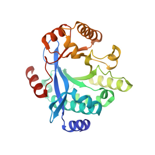
Dimerisation induced formation of the active site and the identification of three metal sites in EAL-phosphodiesterases.

Bellini, D.Horrell, S.Hutchin, A.Phippen, C.W.Strange, R.W.Cai, Y.Wagner, A.Webb, J.S.Tews, I.Walsh, M.A.

(2017) Sci Rep 7: 42166-42166

  • DOI: https://doi.org/10.1038/srep42166
  • Primary Citation Related Structures: 
    4Y9M, 5M1T, 5MF5, 5MFU, 5MKG

  • PubMed Abstract: 

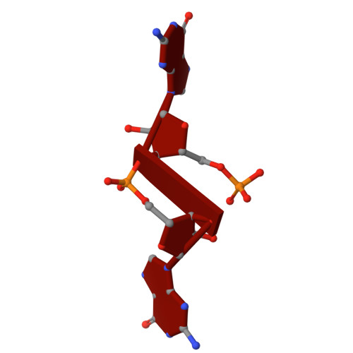
    The bacterial second messenger cyclic di-3',5'-guanosine monophosphate (c-di-GMP) is a key regulator of bacterial motility and virulence. As high levels of c-di-GMP are associated with the biofilm lifestyle, c-di-GMP hydrolysing phosphodiesterases (PDEs) have been identified as key targets to aid development of novel strategies to treat chronic infection by exploiting biofilm dispersal. We have studied the EAL signature motif-containing phosphodiesterase domains from the Pseudomonas aeruginosa proteins PA3825 (PA3825 EAL ) and PA1727 (MucR EAL ). Different dimerisation interfaces allow us to identify interface independent principles of enzyme regulation. Unlike previously characterised two-metal binding EAL-phosphodiesterases, PA3825 EAL in complex with pGpG provides a model for a third metal site. The third metal is positioned to stabilise the negative charge of the 5'-phosphate, and thus three metals could be required for catalysis in analogy to other nucleases. This newly uncovered variation in metal coordination may provide a further level of bacterial PDE regulation.


  • Organizational Affiliation
    • Diamond Light Source, Harwell Science and Innovation Campus, Didcot, Oxfordshire, OX11 0FA, United Kingdom.

Macromolecule Content 

  • Total Structure Weight: 28.95 kDa 
  • Atom Count: 2,209 
  • Modeled Residue Count: 256 
  • Deposited Residue Count: 256 
  • Unique protein chains: 1
  • Unique nucleic acid chains: 1

Macromolecules


Find similar proteins by:|  3D Structure
Entity ID: 1
MoleculeChains  Sequence LengthOrganismDetailsImage
cyclic-guanylate-specific phosphodiesterase254Pseudomonas aeruginosaMutation(s): 0 
Gene Names: PA3825
EC: 3.1.4.52
UniProt
Find proteins for Q9HXH7 (Pseudomonas aeruginosa (strain ATCC 15692 / DSM 22644 / CIP 104116 / JCM 14847 / LMG 12228 / 1C / PRS 101 / PAO1))
Explore Q9HXH7 
Go to UniProtKB:  Q9HXH7
Entity Groups
Sequence Clusters30% Identity50% Identity70% Identity90% Identity95% Identity100% Identity
UniProt GroupQ9HXH7
Sequence Annotations
Expand
Reference Sequence
Find similar nucleic acids by:  Sequence
Entity ID: 2
MoleculeChains LengthOrganismImage
RNA (pGpG)2synthetic construct
Sequence Annotations
Expand
Reference Sequence

Experimental Data & Validation

Experimental Data

  • Method: X-RAY DIFFRACTION
  • Resolution: 2.15 Å
  • R-Value Free:  0.261 (Depositor), 0.255 (DCC) 
  • R-Value Work:  0.202 (Depositor), 0.208 (DCC) 
  • R-Value Observed: 0.205 (Depositor) 
Space Group: P 43 21 2
Unit Cell:
Length ( Å )Angle ( ˚ )
a = 64.78α = 90
b = 64.78β = 90
c = 135.787γ = 90
Software Package:
Software NamePurpose
REFMACrefinement
Aimlessdata scaling
PDB_EXTRACTdata extraction
xia2data reduction
MOLREPphasing

Structure Validation

View Full Validation Report


Entry History 

Deposition Data

Revision History  (Full details and data files)

  • Version 1.0: 2017-03-01
    Type: Initial release
  • Version 1.1: 2019-07-03
    Changes: Data collection, Source and taxonomy
  • Version 1.2: 2019-10-16
    Changes: Data collection
  • Version 1.3: 2024-01-17
    Changes: Data collection, Database references, Derived calculations, Refinement description
  • Version 2.0: 2025-12-17
    Changes: Advisory, Atomic model, Data collection, Database references, Derived calculations, Polymer sequence, Source and taxonomy, Structure summary