5M6J

Crystal structure of nitrophorin 7 E27V mutant from Rhodnius prolixus


Experimental Data Snapshot

  • Method: X-RAY DIFFRACTION
  • Resolution: 1.70 Å
  • R-Value Free: 0.213 
  • R-Value Work: 0.161 
  • R-Value Observed: 0.164 

wwPDB Validation   3D Report Full Report


Ligand Structure Quality Assessment 


This is version 1.2 of the entry. See complete history


Literature

Electrostatic Tuning of the Ligand Binding Mechanism by Glu27 in Nitrophorin 7.

Abbruzzetti, S.Allegri, A.Bidon-Chanal, A.Ogata, H.Soavi, G.Cerullo, G.Bruno, S.Montali, C.Luque, F.J.Viappiani, C.

(2018) Sci Rep 8: 10855-10855

  • DOI: https://doi.org/10.1038/s41598-018-29182-3
  • Primary Citation of Related Structures:  
    5M6J, 5M6K

  • PubMed Abstract: 

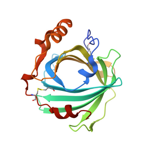
    Nitrophorins (NP) 1-7 are NO-carrying heme proteins found in the saliva of the blood-sucking insect Rhodnius prolixus. The isoform NP7 displays peculiar properties, such as an abnormally high isoelectric point, the ability to bind negatively charged membranes, and a strong pH sensitivity of NO affinity. A unique trait of NP7 is the presence of Glu in position 27, which is occupied by Val in other NPs. Glu27 appears to be important for tuning the heme properties, but its influence on the pH-dependent NO release mechanism, which is assisted by a conformational change in the AB loop, remains unexplored. Here, in order to gain insight into the functional role of Glu27, we examine the effect of Glu27 → Val and Glu27 → Gln mutations on the ligand binding kinetics using CO as a model. The results reveal that annihilation of the negative charge of Glu27 upon mutation reduces the pH sensitivity of the ligand binding rate, a process that in turn depends on the ionization of Asp32. We propose that Glu27 exerts a through-space electrostatic action on Asp32, which shifts the pKa of the latter amino acid towards more acidic values thus reducing the pH sensitivity of the transition between open and closed states.


  • Organizational Affiliation

    Dipartimento di Scienze Matematiche, Fisiche e Informatiche, Università degli Studi di Parma, Parco Area delle Scienze 7/A, 43124, Parma, Italy. stefania.abbruzzetti@unipr.it.


Macromolecules
Find similar proteins by:  (by identity cutoff)  |  3D Structure
Entity ID: 1
MoleculeChains Sequence LengthOrganismDetailsImage
Nitrophorin-7184Rhodnius prolixusMutation(s): 1 
EC: 1.7.6.1
UniProt
Find proteins for Q6PQK2 (Rhodnius prolixus)
Explore Q6PQK2 
Go to UniProtKB:  Q6PQK2
Entity Groups  
Sequence Clusters30% Identity50% Identity70% Identity90% Identity95% Identity100% Identity
UniProt GroupQ6PQK2
Sequence Annotations
Expand
  • Reference Sequence
Small Molecules
Ligands 1 Unique
IDChains Name / Formula / InChI Key2D Diagram3D Interactions
HEM
Query on HEM

Download Ideal Coordinates CCD File 
B [auth A]PROTOPORPHYRIN IX CONTAINING FE
C34 H32 Fe N4 O4
KABFMIBPWCXCRK-RGGAHWMASA-L
Experimental Data & Validation

Experimental Data

  • Method: X-RAY DIFFRACTION
  • Resolution: 1.70 Å
  • R-Value Free: 0.213 
  • R-Value Work: 0.161 
  • R-Value Observed: 0.164 
  • Space Group: P 1 21 1
Unit Cell:
Length ( Å )Angle ( ˚ )
a = 38.228α = 90
b = 66.885β = 116.57
c = 38.714γ = 90
Software Package:
Software NamePurpose
PHENIXrefinement
XDSdata reduction
XSCALEdata scaling
MOLREPphasing

Structure Validation

View Full Validation Report


Ligand Structure Quality Assessment 


Entry History 

Deposition Data

  • Released Date: 2017-12-20 
  • Deposition Author(s): Ogata, H.

Revision History  (Full details and data files)

  • Version 1.0: 2017-12-20
    Type: Initial release
  • Version 1.1: 2018-08-22
    Changes: Data collection, Database references
  • Version 1.2: 2024-01-17
    Changes: Data collection, Database references, Refinement description