5LYU

The native crystal structure of 7SK 5'-hairpin


Experimental Data Snapshot

  • Method: X-RAY DIFFRACTION
  • Resolution: 2.20 Å
  • R-Value Free: 0.240 
  • R-Value Work: 0.191 
  • R-Value Observed: 0.194 

wwPDB Validation   3D Report Full Report


This is version 1.3 of the entry. See complete history


Literature

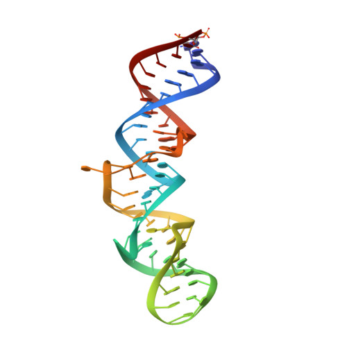
The crystal structure of the 5 functional domain of the transcription riboregulator 7SK.

Martinez-Zapien, D.Legrand, P.McEwen, A.G.Proux, F.Cragnolini, T.Pasquali, S.Dock-Bregeon, A.C.

(2017) Nucleic Acids Res 45: 3568-3579

  • DOI: https://doi.org/10.1093/nar/gkw1351
  • Primary Citation of Related Structures:  
    5LYS, 5LYU, 5LYV

  • PubMed Abstract: 
  • In vertebrates, the 7SK RNA forms the scaffold of a complex, which regulates transcription pausing of RNA-polymerase II. By binding to the HEXIM protein, the complex comprising proteins LARP7 and MePCE captures the positive transcription elongation factor P-TEFb and prevents phosphorylation of pausing factors ...

    In vertebrates, the 7SK RNA forms the scaffold of a complex, which regulates transcription pausing of RNA-polymerase II. By binding to the HEXIM protein, the complex comprising proteins LARP7 and MePCE captures the positive transcription elongation factor P-TEFb and prevents phosphorylation of pausing factors. The HEXIM-binding site embedded in the 5΄-hairpin of 7SK (HP1) encompasses a short signature sequence, a GAUC repeat framed by single-stranded uridines. The present crystal structure of HP1 shows a remarkably straight helical stack involving several unexpected triples formed at a central region. Surprisingly, two uridines of the signature sequence make triple interactions in the major groove of the (GAUC)2. The third uridine is turned outwards or inward, wedging between the other uridines, thus filling the major groove. A molecular dynamics simulation indicates that these two conformations of the signature sequence represent stable alternatives. Analyses of the interaction with the HEXIM protein confirm the importance of the triple interactions at the signature sequence. Altogether, the present structural analysis of 7SK HP1 highlights an original mechanism of swapping bases, which could represent a possible '7SK signature' and provides new insight into the functional importance of the plasticity of RNA.


    Organizational Affiliation

    CNRS, UMR 8227, Integrative Biology of Marine Models, Station Biologique de Roscoff, CS 90074, F-29688, Roscoff cedex, France.


Macromolecules
Find similar nucleic acids by:  (by identity cutoff)  |  3D Structure
Entity ID: 1
MoleculeChainsLengthOrganismImage
7SK RNA
A, B
58Homo sapiens
Protein Feature View
Expand
  • Reference Sequence
Small Molecules
Ligands 3 Unique
IDChainsName / Formula / InChI Key2D Diagram3D Interactions
CL
Query on CL

Download Ideal Coordinates CCD File 
D [auth A],
F [auth B]
CHLORIDE ION
Cl
VEXZGXHMUGYJMC-UHFFFAOYSA-M
 Ligand Interaction
MG
Query on MG

Download Ideal Coordinates CCD File 
E [auth B]MAGNESIUM ION
Mg
JLVVSXFLKOJNIY-UHFFFAOYSA-N
 Ligand Interaction
NA
Query on NA

Download Ideal Coordinates CCD File 
C [auth A]SODIUM ION
Na
FKNQFGJONOIPTF-UHFFFAOYSA-N
 Ligand Interaction
Experimental Data & Validation

Experimental Data

  • Method: X-RAY DIFFRACTION
  • Resolution: 2.20 Å
  • R-Value Free: 0.240 
  • R-Value Work: 0.191 
  • R-Value Observed: 0.194 
  • Space Group: P 1 21 1
Unit Cell:
Length ( Å )Angle ( ˚ )
a = 45.79α = 90
b = 47.78β = 99.05
c = 68.67γ = 90
Software Package:
Software NamePurpose
XSCALEdata scaling
BUSTER-TNTrefinement
PDB_EXTRACTdata extraction
XDSdata reduction
PHASERphasing

Structure Validation

View Full Validation Report


Entry History & Funding Information

Deposition Data


Funding OrganizationLocationGrant Number
French National Research AgencyFranceANR-06-BLAN-0072
French National Research AgencyFranceANR-12-BSV5-0018
French National Research AgencyFranceANR-10-INSB-05-01

Revision History  (Full details and data files)

  • Version 1.0: 2017-01-25
    Type: Initial release
  • Version 1.1: 2017-05-03
    Changes: Database references
  • Version 1.2: 2017-08-30
    Changes: Author supporting evidence, Derived calculations
  • Version 1.3: 2018-04-18
    Changes: Data collection, Structure summary