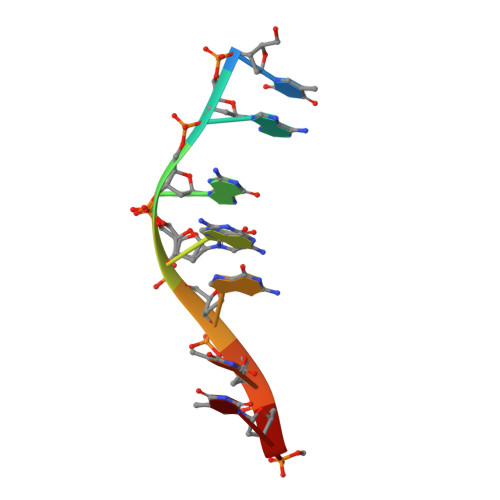
5LS8

Light-activated ruthenium complex bound to a DNA quadruplex


Experimental Data Snapshot

  • Method: X-RAY DIFFRACTION
  • Resolution: 1.78 Å
  • R-Value Free: 0.233 
  • R-Value Work: 0.214 
  • R-Value Observed: 0.215 

wwPDB Validation   3D Report Full Report


Ligand Structure Quality Assessment 


This is version 3.1 of the entry. See complete history


Literature

Structural Studies Reveal Enantiospecific Recognition of a DNA G-Quadruplex by a Ruthenium Polypyridyl Complex.

McQuaid, K.Abell, H.Gurung, S.P.Allan, D.R.Winter, G.Sorensen, T.Cardin, D.J.Brazier, J.A.Cardin, C.J.Hall, J.P.

(2019) Angew Chem Int Ed Engl 58: 9881-9885

  • DOI: https://doi.org/10.1002/anie.201814502
  • Primary Citation of Related Structures:  
    5LS8

  • PubMed Abstract: 

    By using X-ray crystallography, we show that the complexes Λ/Δ-[Ru(TAP) 2 (11-CN-dppz)] 2+ (TAP=1,4,5,8-tetraazaphenanthrene, dppz=dipyridophenazine) bind DNA G-quadruplex in an enantiospecific manner that parallels the specificity of these complexes with duplex DNA. The Λ complex crystallises with the normally parallel stranded d(TAGGGTTA) tetraplex to give the first such antiparallel strand assembly in which syn-guanosine is adjacent to the complex at the 5' end of the quadruplex core. SRCD measurements confirm that the same conformational switch occurs in solution. The Δ enantiomer, by contrast, is present in the structure but stacked at the ends of the assembly. In addition, we report the structure of Λ-[Ru(phen) 2 (11-CN-dppz)] 2+ bound to d(TCGGCGCCGA), a duplex-forming sequence, and use both structural models to provide insight into the motif-specific luminescence response of the isostructural phen analogue enantiomers.


  • Organizational Affiliation

    Department of Chemistry, University of Reading, Whiteknights, Reading, RG6 6AD, UK.


Macromolecules

Find similar nucleic acids by:  Sequence   |   3D Structure  

Entity ID: 1
MoleculeChains LengthOrganismImage
DNA (5'-D(*TP*AP*GP*GP*GP*TP*TP*A)-3')
A, B, C, D
8synthetic construct
Sequence Annotations
Expand
  • Reference Sequence
Small Molecules
Ligands 3 Unique
IDChains Name / Formula / InChI Key2D Diagram3D Interactions
QHL
Query on QHL

Download Ideal Coordinates CCD File 
F [auth A],
K [auth C]
delta-[Ru(tap2-dppz-CN)]2+
C39 H21 N13 Ru
HNFPSJMHTKCUIN-UHFFFAOYSA-N
KSB
Query on KSB

Download Ideal Coordinates CCD File 
E [auth A],
I [auth B],
J [auth C],
L [auth D]
lambda-[Ru(tap2-dppz-CN)]2+
C39 H21 N13 Ru
HNFPSJMHTKCUIN-UHFFFAOYSA-N
K
Query on K

Download Ideal Coordinates CCD File 
G [auth A],
H [auth A]
POTASSIUM ION
K
NPYPAHLBTDXSSS-UHFFFAOYSA-N
Experimental Data & Validation

Experimental Data

  • Method: X-RAY DIFFRACTION
  • Resolution: 1.78 Å
  • R-Value Free: 0.233 
  • R-Value Work: 0.214 
  • R-Value Observed: 0.215 
  • Space Group: P 65
Unit Cell:
Length ( Å )Angle ( ˚ )
a = 36.625α = 90
b = 36.625β = 90
c = 136.104γ = 120
Software Package:
Software NamePurpose
PHENIXrefinement
DIALSdata reduction
DIALSdata scaling
SHELXDEphasing
xia2data scaling

Structure Validation

View Full Validation Report


Ligand Structure Quality Assessment 


Entry History & Funding Information

Deposition Data


Funding OrganizationLocationGrant Number
Biotechnology and Biological Sciences Research CouncilUnited KingdomBB/K019279/1
Biotechnology and Biological Sciences Research CouncilUnited KingdomMM/M004635/1

Revision History  (Full details and data files)

  • Version 1.0: 2018-01-17
    Type: Initial release
  • Version 2.0: 2018-10-03
    Changes: Atomic model, Data collection, Derived calculations, Non-polymer description, Structure summary
  • Version 2.1: 2019-08-28
    Changes: Data collection, Database references
  • Version 2.2: 2019-09-11
    Changes: Data collection, Database references
  • Version 3.0: 2023-12-27
    Changes: Atomic model, Data collection, Database references, Derived calculations, Non-polymer description, Structure summary
  • Version 3.1: 2024-01-10
    Changes: Derived calculations, Structure summary