5LS8 | pdb_00005ls8

Light-activated ruthenium complex bound to a DNA quadruplex


Experimental Data Snapshot

  • Method: X-RAY DIFFRACTION
  • Resolution: 1.78 Å
  • R-Value Free: 
    0.233 (Depositor), 0.236 (DCC) 
  • R-Value Work: 
    0.214 (Depositor), 0.217 (DCC) 
  • R-Value Observed: 
    0.215 (Depositor) 

wwPDB Validation 3D Report Full Report

Validation slider image for 5LS8

Ligand Structure Quality Assessment 


This is version 3.1 of the entry. See complete history

Literature

Structural Studies Reveal Enantiospecific Recognition of a DNA G-Quadruplex by a Ruthenium Polypyridyl Complex.

McQuaid, K.Abell, H.Gurung, S.P.Allan, D.R.Winter, G.Sorensen, T.Cardin, D.J.Brazier, J.A.Cardin, C.J.Hall, J.P.

(2019) Angew Chem Int Ed Engl 58: 9881-9885

  • DOI: https://doi.org/10.1002/anie.201814502
  • Primary Citation Related Structures: 
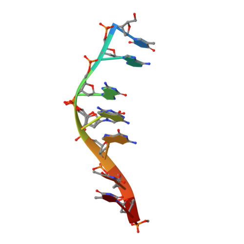
    5LS8

  • PubMed Abstract: 

    By using X-ray crystallography, we show that the complexes Λ/Δ-[Ru(TAP) 2 (11-CN-dppz)] 2+ (TAP=1,4,5,8-tetraazaphenanthrene, dppz=dipyridophenazine) bind DNA G-quadruplex in an enantiospecific manner that parallels the specificity of these complexes with duplex DNA. The Λ complex crystallises with the normally parallel stranded d(TAGGGTTA) tetraplex to give the first such antiparallel strand assembly in which syn-guanosine is adjacent to the complex at the 5' end of the quadruplex core. SRCD measurements confirm that the same conformational switch occurs in solution. The Δ enantiomer, by contrast, is present in the structure but stacked at the ends of the assembly. In addition, we report the structure of Λ-[Ru(phen) 2 (11-CN-dppz)] 2+ bound to d(TCGGCGCCGA), a duplex-forming sequence, and use both structural models to provide insight into the motif-specific luminescence response of the isostructural phen analogue enantiomers.


  • Organizational Affiliation
    • Department of Chemistry, University of Reading, Whiteknights, Reading, RG6 6AD, UK.

Macromolecule Content 

  • Total Structure Weight: 14.64 kDa 
  • Atom Count: 1,043 
  • Modeled Residue Count: 32 
  • Deposited Residue Count: 32 
  • Unique nucleic acid chains: 1

Macromolecules

Find similar nucleic acids by:  Sequence
Entity ID: 1
MoleculeChains LengthOrganismImage
DNA (5'-D(*TP*AP*GP*GP*GP*TP*TP*A)-3')
A, B, C, D
8synthetic construct
Sequence Annotations
Expand
Reference Sequence

Small Molecules

Ligands 3 Unique
IDChains Name / Formula / InChI Key2D Diagram3D Interactions
KSB

Query on KSB


Download:Ideal Coordinates CCD File
E [auth A],
I [auth B],
J [auth C],
L [auth D]
lambda-[Ru(tap2-dppz-CN)]2+
C39 H21 N13 Ru
HNFPSJMHTKCUIN-UHFFFAOYSA-N
QHL

Query on QHL


Download:Ideal Coordinates CCD File
F [auth A],
K [auth C]
delta-[Ru(tap2-dppz-CN)]2+
C39 H21 N13 Ru
HNFPSJMHTKCUIN-UHFFFAOYSA-N
K

Query on K


Download:Ideal Coordinates CCD File
G [auth A],
H [auth A]
POTASSIUM ION
K
NPYPAHLBTDXSSS-UHFFFAOYSA-N

Experimental Data & Validation

Experimental Data

  • Method: X-RAY DIFFRACTION
  • Resolution: 1.78 Å
  • R-Value Free:  0.233 (Depositor), 0.236 (DCC) 
  • R-Value Work:  0.214 (Depositor), 0.217 (DCC) 
  • R-Value Observed: 0.215 (Depositor) 
Space Group: P 65
Unit Cell:
Length ( Å )Angle ( ˚ )
a = 36.625α = 90
b = 36.625β = 90
c = 136.104γ = 120
Software Package:
Software NamePurpose
PHENIXrefinement
DIALSdata reduction
DIALSdata scaling
SHELXDEphasing
xia2data scaling

Structure Validation

View Full Validation Report


Ligand Structure Quality Assessment 


Entry History 

& Funding Information

Deposition Data


Funding OrganizationLocationGrant Number
Biotechnology and Biological Sciences Research CouncilUnited KingdomBB/K019279/1
Biotechnology and Biological Sciences Research CouncilUnited KingdomMM/M004635/1

Revision History  (Full details and data files)

  • Version 1.0: 2018-01-17
    Type: Initial release
  • Version 2.0: 2018-10-03
    Changes: Atomic model, Data collection, Derived calculations, Non-polymer description, Structure summary
  • Version 2.1: 2019-08-28
    Changes: Data collection, Database references
  • Version 2.2: 2019-09-11
    Changes: Data collection, Database references
  • Version 3.0: 2023-12-27
    Changes: Atomic model, Data collection, Database references, Derived calculations, Non-polymer description, Structure summary
  • Version 3.1: 2024-01-10
    Changes: Derived calculations, Structure summary