5LJN

Structure of the HOIP PUB domain bound to SPATA2 PIM peptide


Experimental Data Snapshot

  • Method: X-RAY DIFFRACTION
  • Resolution: 2.70 Å
  • R-Value Free: 0.281 
  • R-Value Work: 0.233 
  • R-Value Observed: 0.235 

wwPDB Validation   3D Report Full Report


This is version 1.3 of the entry. See complete history


Literature

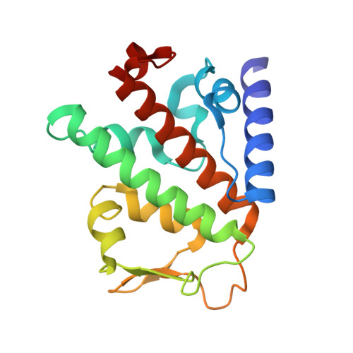
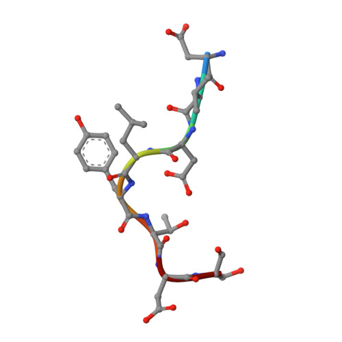
SPATA2 Links CYLD to LUBAC, Activates CYLD, and Controls LUBAC Signaling.

Elliott, P.R.Leske, D.Hrdinka, M.Bagola, K.Fiil, B.K.McLaughlin, S.H.Wagstaff, J.Volkmar, N.Christianson, J.C.Kessler, B.M.Freund, S.M.Komander, D.Gyrd-Hansen, M.

(2016) Mol Cell 63: 990-1005

  • DOI: https://doi.org/10.1016/j.molcel.2016.08.001
  • Primary Citation of Related Structures:  
    5LJM, 5LJN

  • PubMed Abstract: 

    The linear ubiquitin chain assembly complex (LUBAC) regulates immune signaling, and its function is regulated by the deubiquitinases OTULIN and CYLD, which associate with the catalytic subunit HOIP. However, the mechanism through which CYLD interacts with HOIP is unclear. We here show that CYLD interacts with HOIP via spermatogenesis-associated protein 2 (SPATA2). SPATA2 interacts with CYLD through its non-canonical PUB domain, which binds the catalytic CYLD USP domain in a CYLD B-box-dependent manner. Significantly, SPATA2 binding activates CYLD-mediated hydrolysis of ubiquitin chains. SPATA2 also harbors a conserved PUB-interacting motif that selectively docks into the HOIP PUB domain. In cells, SPATA2 is recruited to the TNF receptor 1 signaling complex and is required for CYLD recruitment. Loss of SPATA2 increases ubiquitination of LUBAC substrates and results in enhanced NOD2 signaling. Our data reveal SPATA2 as a high-affinity binding partner of CYLD and HOIP, and a regulatory component of LUBAC-mediated NF-κB signaling.


  • Organizational Affiliation

    Division of Protein and Nucleic Acid Chemistry, MRC Laboratory of Molecular Biology, Francis Crick Avenue, Cambridge CB2 0QH, UK.


Macromolecules
Find similar proteins by:  (by identity cutoff)  |  3D Structure
Entity ID: 1
MoleculeChains Sequence LengthOrganismDetailsImage
E3 ubiquitin-protein ligase RNF31
A, B
173Homo sapiensMutation(s): 0 
Gene Names: RNF31ZIBRA
EC: 6.3.2
UniProt & NIH Common Fund Data Resources
Find proteins for Q96EP0 (Homo sapiens)
Explore Q96EP0 
Go to UniProtKB:  Q96EP0
PHAROS:  Q96EP0
GTEx:  ENSG00000092098 
Entity Groups  
Sequence Clusters30% Identity50% Identity70% Identity90% Identity95% Identity100% Identity
UniProt GroupQ96EP0
Sequence Annotations
Expand
  • Reference Sequence

Find similar proteins by:  Sequence   |   3D Structure  

Entity ID: 2
MoleculeChains Sequence LengthOrganismDetailsImage
Spermatogenesis-associated protein 2
C, D
8Homo sapiensMutation(s): 0 
UniProt & NIH Common Fund Data Resources
Find proteins for Q9UM82 (Homo sapiens)
Explore Q9UM82 
Go to UniProtKB:  Q9UM82
PHAROS:  Q9UM82
GTEx:  ENSG00000158480 
Entity Groups  
UniProt GroupQ9UM82
Sequence Annotations
Expand
  • Reference Sequence
Experimental Data & Validation

Experimental Data

  • Method: X-RAY DIFFRACTION
  • Resolution: 2.70 Å
  • R-Value Free: 0.281 
  • R-Value Work: 0.233 
  • R-Value Observed: 0.235 
  • Space Group: P 43
Unit Cell:
Length ( Å )Angle ( ˚ )
a = 89.04α = 90
b = 89.04β = 90
c = 53.563γ = 90
Software Package:
Software NamePurpose
Aimlessdata scaling
PHENIXrefinement
PDB_EXTRACTdata extraction
DIALSdata reduction
PHASERphasing

Structure Validation

View Full Validation Report


Entry History 

Deposition Data

Revision History  (Full details and data files)

  • Version 1.0: 2016-08-24
    Type: Initial release
  • Version 1.1: 2016-09-14
    Changes: Database references
  • Version 1.2: 2016-09-28
    Changes: Database references
  • Version 1.3: 2024-01-10
    Changes: Data collection, Database references, Refinement description