Uncoupling conformational states from activity in an allosteric enzyme.
Pisco, J.P., Chiara, C., Pacholarz, K.J., Garza-Garcia, A., Ogrodowicz, R.W., Walker, P.A., Barran, P.E., Smerdon, S.J., Carvalho, L.P.S.(2017) Nat Commun 8: 203-203
- PubMed: 28781362 Search on PubMedSearch on PubMed Central
- DOI: https://doi.org/10.1038/s41467-017-00224-0
- Primary Citation Related Structures:
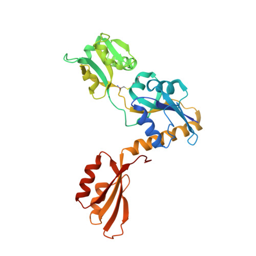
5LHT, 5LHU - PubMed Abstract:
ATP-phosphoribosyltransferase (ATP-PRT) is a hexameric enzyme in conformational equilibrium between an open and seemingly active state and a closed and presumably inhibited form. The structure-function relationship of allosteric regulation in this system is still not fully understood. Here, we develop a screening strategy for modulators of ATP-PRT and identify 3-(2-thienyl)-L-alanine (TIH) as an allosteric activator of this enzyme. Kinetic analysis reveals co-occupancy of the allosteric sites by TIH and L-histidine. Crystallographic and native ion-mobility mass spectrometry data show that the TIH-bound activated form of the enzyme closely resembles the inhibited L-histidine-bound closed conformation, revealing the uncoupling between ATP-PRT open and closed conformations and its functional state. These findings suggest that dynamic processes are responsible for ATP-PRT allosteric regulation and that similar mechanisms might also be found in other enzymes bearing a ferredoxin-like allosteric domain.Active and inactive state ATP-phosphoribosyltransferases (ATP-PRTs) are believed to have different conformations. Here the authors show that in both states, ATP-PRT has a similar structural arrangement, suggesting that dynamic alterations are involved in ATP-PRT regulation by allosteric modulators.
- Mycobacterial Metabolism and Antibiotic Research Laboratory, The Francis Crick Institute, 1 Midland Road, London, NW1 1AT, UK.
Organizational Affiliation: