5LA1

The mechanism by which arabinoxylanases can recognise highly decorated xylans


Experimental Data Snapshot

  • Method: X-RAY DIFFRACTION
  • Resolution: 1.90 Å
  • R-Value Free: 0.160 
  • R-Value Work: 0.129 
  • R-Value Observed: 0.131 

wwPDB Validation   3D Report Full Report


Ligand Structure Quality Assessment 


This is version 3.1 of the entry. See complete history


Literature

The Mechanism by Which Arabinoxylanases Can Recognize Highly Decorated Xylans.

Labourel, A.Crouch, L.I.Bras, J.L.Jackson, A.Rogowski, A.Gray, J.Yadav, M.P.Henrissat, B.Fontes, C.M.Gilbert, H.J.Najmudin, S.Basle, A.Cuskin, F.

(2016) J Biol Chem 291: 22149-22159

  • DOI: https://doi.org/10.1074/jbc.M116.743948
  • Primary Citation of Related Structures:  
    5G56, 5LA0, 5LA1, 5LA2

  • PubMed Abstract: 

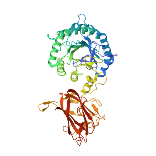
    The enzymatic degradation of plant cell walls is an important biological process of increasing environmental and industrial significance. Xylan, a major component of the plant cell wall, consists of a backbone of β-1,4-xylose (Xylp) units that are often decorated with arabinofuranose (Araf) side chains. A large penta-modular enzyme, CtXyl5A, was shown previously to specifically target arabinoxylans. The mechanism of substrate recognition displayed by the enzyme, however, remains unclear. Here we report the crystal structure of the arabinoxylanase and the enzyme in complex with ligands. The data showed that four of the protein modules adopt a rigid structure, which stabilizes the catalytic domain. The C-terminal non-catalytic carbohydrate binding module could not be observed in the crystal structure, suggesting positional flexibility. The structure of the enzyme in complex with Xylp-β-1,4-Xylp-β-1,4-Xylp-[α-1,3-Araf]-β-1,4-Xylp showed that the Araf decoration linked O 3 to the xylose in the active site is located in the pocket (-2* subsite) that abuts onto the catalytic center. The -2* subsite can also bind to Xylp and Arap, explaining why the enzyme can utilize xylose and arabinose as specificity determinants. Alanine substitution of Glu 68 , Tyr 92 , or Asn 139 , which interact with arabinose and xylose side chains at the -2* subsite, abrogates catalytic activity. Distal to the active site, the xylan backbone makes limited apolar contacts with the enzyme, and the hydroxyls are solvent-exposed. This explains why CtXyl5A is capable of hydrolyzing xylans that are extensively decorated and that are recalcitrant to classic endo-xylanase attack.


  • Organizational Affiliation

    From the Institute for Cell and Molecular Biosciences, Newcastle University, Newcastle upon Tyne NE2 4HH, United Kingdom.


Macromolecules
Find similar proteins by:  (by identity cutoff)  |  3D Structure
Entity ID: 1
MoleculeChains Sequence LengthOrganismDetailsImage
Carbohydrate binding family 6491Acetivibrio thermocellus JW20Mutation(s): 0 
Gene Names: Cther_1146
UniProt
Find proteins for A3DHG6 (Acetivibrio thermocellus (strain ATCC 27405 / DSM 1237 / JCM 9322 / NBRC 103400 / NCIMB 10682 / NRRL B-4536 / VPI 7372))
Explore A3DHG6 
Go to UniProtKB:  A3DHG6
Entity Groups  
Sequence Clusters30% Identity50% Identity70% Identity90% Identity95% Identity100% Identity
UniProt GroupA3DHG6
Sequence Annotations
Expand
  • Reference Sequence
Small Molecules
Ligands 3 Unique
IDChains Name / Formula / InChI Key2D Diagram3D Interactions
XYP
Query on XYP

Download Ideal Coordinates CCD File 
B [auth A],
C [auth A]
beta-D-xylopyranose
C5 H10 O5
SRBFZHDQGSBBOR-KKQCNMDGSA-N
144
Query on 144

Download Ideal Coordinates CCD File 
G [auth A],
H [auth A],
I [auth A]
TRIS-HYDROXYMETHYL-METHYL-AMMONIUM
C4 H12 N O3
DRDCQJADRSJFFD-UHFFFAOYSA-N
CA
Query on CA

Download Ideal Coordinates CCD File 
D [auth A],
E [auth A],
F [auth A]
CALCIUM ION
Ca
BHPQYMZQTOCNFJ-UHFFFAOYSA-N
Experimental Data & Validation

Experimental Data

  • Method: X-RAY DIFFRACTION
  • Resolution: 1.90 Å
  • R-Value Free: 0.160 
  • R-Value Work: 0.129 
  • R-Value Observed: 0.131 
  • Space Group: P 21 21 21
Unit Cell:
Length ( Å )Angle ( ˚ )
a = 67.911α = 90
b = 72.514β = 90
c = 109.537γ = 90
Software Package:
Software NamePurpose
REFMACrefinement
Aimlessdata scaling
MOLREPphasing
XDSdata reduction

Structure Validation

View Full Validation Report


Ligand Structure Quality Assessment 


Entry History & Funding Information

Deposition Data


Funding OrganizationLocationGrant Number
Biotechnology and Biological Sciences Research CouncilUnited KingdomBB/K020358/1

Revision History  (Full details and data files)

  • Version 1.0: 2016-08-31
    Type: Initial release
  • Version 1.1: 2016-10-26
    Changes: Database references
  • Version 1.2: 2017-08-30
    Changes: Author supporting evidence
  • Version 2.0: 2018-10-24
    Changes: Advisory, Atomic model, Data collection, Derived calculations, Non-polymer description, Structure summary
  • Version 3.0: 2020-07-29
    Type: Remediation
    Reason: Carbohydrate remediation
    Changes: Atomic model, Data collection, Derived calculations, Structure summary
  • Version 3.1: 2024-01-10
    Changes: Data collection, Database references, Refinement description, Structure summary