5KOJ | pdb_00005koj

Nitrogenase MoFeP protein in the IDS oxidized state


Experimental Data Snapshot

  • Method: X-RAY DIFFRACTION
  • Resolution: 2.59 Å
  • R-Value Free: 
    0.208 (Depositor), 0.210 (DCC) 
  • R-Value Work: 
    0.168 (Depositor), 0.175 (DCC) 
  • R-Value Observed: 
    0.170 (Depositor) 

wwPDB Validation 3D Report Full Report

Validation slider image for 5KOJ

Ligand Structure Quality Assessment 


This is version 1.2 of the entry. See complete history

Literature

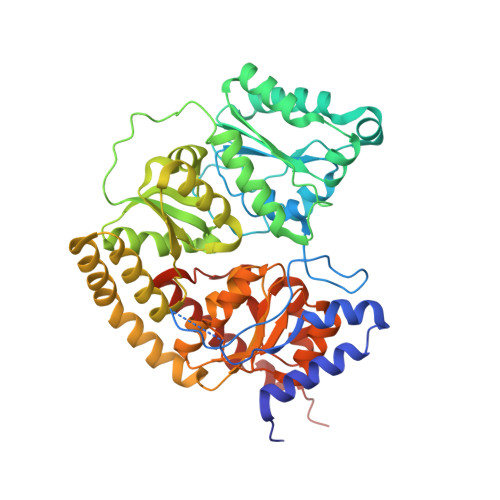
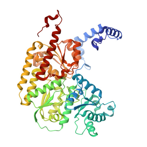
Tyrosine-Coordinated P-Cluster in G. diazotrophicus Nitrogenase: Evidence for the Importance of O-Based Ligands in Conformationally Gated Electron Transfer.

Owens, C.P.Katz, F.E.Carter, C.H.Oswald, V.F.Tezcan, F.A.

(2016) J Am Chem Soc 138: 10124-10127

  • DOI: https://doi.org/10.1021/jacs.6b06783
  • Primary Citation Related Structures: 
    5KOH, 5KOJ

  • PubMed Abstract: 

    The P-cluster is a unique iron-sulfur center that likely functions as a dynamic electron (e(-)) relay site between the Fe-protein and the catalytic FeMo-cofactor in nitrogenase. The P-cluster has been shown to undergo large conformational changes upon 2-e(-) oxidation which entail the coordination of two of the Fe centers to a Ser side chain and a backbone amide N, respectively. Yet, how and if this 2-e(-) oxidized state (P(OX)) is involved in catalysis by nitrogenase is not well established. Here, we present the crystal structures of reduced and oxidized MoFe-protein (MoFeP) from Gluconacetobacter diazotrophicus (Gd), which natively possesses an Ala residue in the position of the Ser ligand to the P-cluster. While reduced Gd-MoFeP is structurally identical to previously characterized counterparts around the FeMo-cofactor, oxidized Gd-MoFeP features an unusual Tyr coordination to its P-cluster along with ligation by a backbone amide nitrogen. EPR analysis of the oxidized Gd-MoFeP P-cluster confirmed that it is a 2-e(-) oxidized, integer-spin species. Importantly, we have found that the sequence positions corresponding to the Ser and Tyr ligands are almost completely covariant among Group I nitrogenases. These findings strongly support the possibility that the P(OX) state is functionally relevant in nitrogenase catalysis and that a hard, O-based anionic ligand serves to stabilize this state in a switchable fashion.


  • Organizational Affiliation
    • Department of Chemistry and Biochemistry, University of California, San Diego , 9500 Gilman Drive, La Jolla, California 92093-0356, United States.

Macromolecule Content 

  • Total Structure Weight: 229.82 kDa 
  • Atom Count: 16,070 
  • Modeled Residue Count: 1,981 
  • Deposited Residue Count: 2,020 
  • Unique protein chains: 2

Macromolecules

Find similar proteins by:|  3D Structure
Entity ID: 1
MoleculeChains  Sequence LengthOrganismDetailsImage
Nitrogenase protein alpha chain
A, C
499Gluconacetobacter diazotrophicus PA1 5Mutation(s): 0 
EC: 1.18.6.1
UniProt
Find proteins for A9H5W5 (Gluconacetobacter diazotrophicus (strain ATCC 49037 / DSM 5601 / CCUG 37298 / CIP 103539 / LMG 7603 / PAl5))
Explore A9H5W5 
Go to UniProtKB:  A9H5W5
Entity Groups
Sequence Clusters30% Identity50% Identity70% Identity90% Identity95% Identity100% Identity
UniProt GroupA9H5W5
Sequence Annotations
Expand
Reference Sequence
Find similar proteins by:|  3D Structure
Entity ID: 2
MoleculeChains  Sequence LengthOrganismDetailsImage
Nitrogenase FeMo beta subunit protein NifK
B, D
511Gluconacetobacter diazotrophicus PA1 5Mutation(s): 0 
EC: 1.18.6.1
UniProt
Find proteins for A9H5W8 (Gluconacetobacter diazotrophicus (strain ATCC 49037 / DSM 5601 / CCUG 37298 / CIP 103539 / LMG 7603 / PAl5))
Explore A9H5W8 
Go to UniProtKB:  A9H5W8
Entity Groups
Sequence Clusters30% Identity50% Identity70% Identity90% Identity95% Identity100% Identity
UniProt GroupA9H5W8
Sequence Annotations
Expand
Reference Sequence

Small Molecules

Ligands 4 Unique
IDChains Name / Formula / InChI Key2D Diagram3D Interactions
ICS

Query on ICS


Download:Ideal Coordinates CCD File
F [auth A],
J [auth C]
iron-sulfur-molybdenum cluster with interstitial carbon
C Fe7 Mo S9
DDQFAOMIVKLFON-UHFFFAOYSA-N
CLF

Query on CLF


Download:Ideal Coordinates CCD File
G [auth A],
K [auth C]
FE(8)-S(7) CLUSTER
Fe8 S7
JKVMXLBGZBULKV-UHFFFAOYSA-N
HCA

Query on HCA


Download:Ideal Coordinates CCD File
E [auth A],
I [auth C]
3-HYDROXY-3-CARBOXY-ADIPIC ACID
C7 H10 O7
XKJVEVRQMLKSMO-SSDOTTSWSA-N
FE

Query on FE


Download:Ideal Coordinates CCD File
H [auth B],
L [auth D]
FE (III) ION
Fe
VTLYFUHAOXGGBS-UHFFFAOYSA-N

Experimental Data & Validation

Experimental Data

  • Method: X-RAY DIFFRACTION
  • Resolution: 2.59 Å
  • R-Value Free:  0.208 (Depositor), 0.210 (DCC) 
  • R-Value Work:  0.168 (Depositor), 0.175 (DCC) 
  • R-Value Observed: 0.170 (Depositor) 
Space Group: P 41 21 2
Unit Cell:
Length ( Å )Angle ( ˚ )
a = 201.569α = 90
b = 201.569β = 90
c = 132.208γ = 90
Software Package:
Software NamePurpose
REFMACrefinement
XDSdata scaling
Cootmodel building
PHASERphasing
Aimlessdata scaling

Structure Validation

View Full Validation Report


Ligand Structure Quality Assessment 


Entry History 

Deposition Data

Revision History  (Full details and data files)

  • Version 1.0: 2016-09-14
    Type: Initial release
  • Version 1.1: 2017-11-22
    Changes: Derived calculations, Refinement description
  • Version 1.2: 2024-03-06
    Changes: Data collection, Database references, Derived calculations, Refinement description