5K7C

The native structure of native pistol ribozyme


Experimental Data Snapshot

  • Method: X-RAY DIFFRACTION
  • Resolution: 2.73 Å
  • R-Value Free: 0.250 
  • R-Value Work: 0.200 

wwPDB Validation   3D Report Full Report


This is version 1.2 of the entry. See complete history


Literature

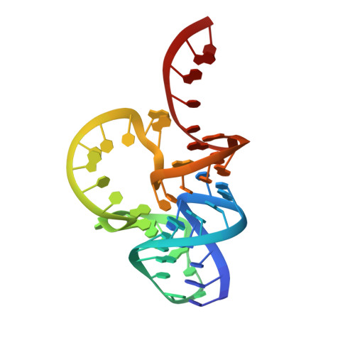
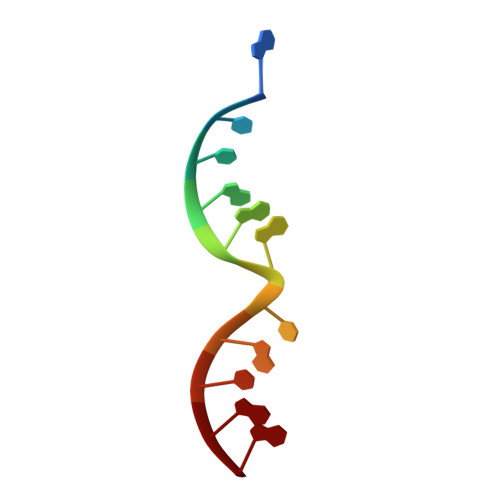
Pistol ribozyme adopts a pseudoknot fold facilitating site-specific in-line cleavage.

Ren, A.Vusurovic, N.Gebetsberger, J.Gao, P.Juen, M.Kreutz, C.Micura, R.Patel, D.J.

(2016) Nat Chem Biol 12: 702-708

  • DOI: https://doi.org/10.1038/nchembio.2125
  • Primary Citation of Related Structures:  
    5K7C, 5K7D, 5K7E

  • PubMed Abstract: 
  • The field of small self-cleaving nucleolytic ribozymes has been invigorated by the recent discovery of the twister, twister-sister, pistol and hatchet ribozymes. We report the crystal structure of a pistol ribozyme termed env25, which adopts a compact tertiary architecture stabilized by an embedded pseudoknot fold ...

    The field of small self-cleaving nucleolytic ribozymes has been invigorated by the recent discovery of the twister, twister-sister, pistol and hatchet ribozymes. We report the crystal structure of a pistol ribozyme termed env25, which adopts a compact tertiary architecture stabilized by an embedded pseudoknot fold. The G-U cleavage site adopts a splayed-apart conformation with in-line alignment of the modeled 2'-O of G for attack on the adjacent to-be-cleaved P-O5' bond. Highly conserved residues G40 (N1 position) and A32 (N3 and 2'-OH positions) are aligned to act as a general base and a general acid, respectively, to accelerate cleavage chemistry, with their roles confirmed by cleavage assays on variants, and an increased pKa of 4.7 for A32. Our structure of the pistol ribozyme defined how the overall and local topologies dictate the in-line alignment at the G-U cleavage site, with cleavage assays on variants revealing key residues that participate in acid-base-catalyzed cleavage chemistry.


    Organizational Affiliation

    Structural Biology Program, Memorial Sloan-Kettering Cancer, New York, New York, USA.


Macromolecules
Find similar nucleic acids by: 3D Structure
Entity ID: 1
MoleculeChainsLengthOrganismImage
RNA 47-MER47synthetic construct
Protein Feature View
Expand
  • Reference Sequence

Find similar nucleic acids by:  Sequence   |   3D Structure  

Entity ID: 2
MoleculeChainsLengthOrganismImage
DNA/RNA 11-MER11synthetic construct
Protein Feature View
Expand
  • Reference Sequence
Small Molecules
Ligands 1 Unique
IDChainsName / Formula / InChI Key2D Diagram3D Interactions
MG
Query on MG

Download Ideal Coordinates CCD File 
C [auth A],
D [auth A],
E [auth A]
MAGNESIUM ION
Mg
JLVVSXFLKOJNIY-UHFFFAOYSA-N
 Ligand Interaction
Experimental Data & Validation

Experimental Data

  • Method: X-RAY DIFFRACTION
  • Resolution: 2.73 Å
  • R-Value Free: 0.250 
  • R-Value Work: 0.200 
  • Space Group: P 32 2 1
Unit Cell:
Length ( Å )Angle ( ˚ )
a = 56.196α = 90
b = 56.196β = 90
c = 89.311γ = 120
Software Package:
Software NamePurpose
PHENIXrefinement
XDSdata reduction
XDSdata scaling
AutoSolphasing

Structure Validation

View Full Validation Report


Entry History 

Deposition Data

Revision History  (Full details and data files)

  • Version 1.0: 2016-07-13
    Type: Initial release
  • Version 1.1: 2016-07-27
    Changes: Database references
  • Version 1.2: 2016-08-31
    Changes: Database references