5K6K | pdb_00005k6k

Zika virus non-structural protein 1 (NS1)


Experimental Data Snapshot

  • Method: X-RAY DIFFRACTION
  • Resolution: 1.89 Å
  • R-Value Free: 
    0.189 (Depositor), 0.190 (DCC) 
  • R-Value Work: 
    0.149 (Depositor), 0.153 (DCC) 
  • R-Value Observed: 
    0.150 (Depositor) 

Starting Model: experimental
View more details

wwPDB Validation 3D Report Full Report

Validation slider image for 5K6K

Ligand Structure Quality Assessment 


This is version 1.7 of the entry. See complete history

Literature

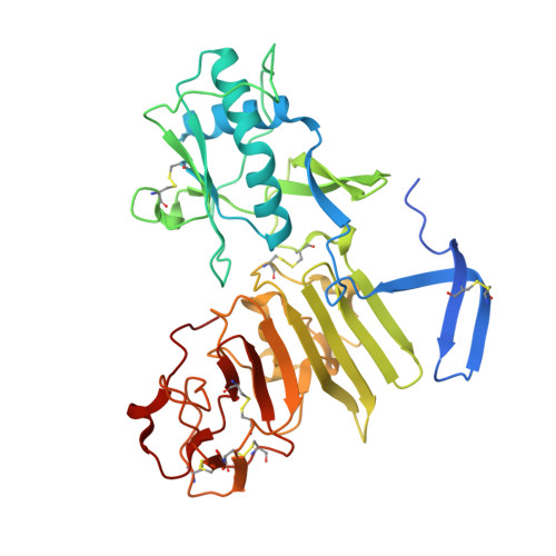
Extended surface for membrane association in Zika virus NS1 structure.

Brown, W.C.Akey, D.L.Konwerski, J.R.Tarrasch, J.T.Skiniotis, G.Kuhn, R.J.Smith, J.L.

(2016) Nat Struct Mol Biol 23: 865-867

  • DOI: https://doi.org/10.1038/nsmb.3268
  • Primary Citation Related Structures: 
    5K6K

  • PubMed Abstract: 

    The Zika virus, which has been implicated in an increase in neonatal microcephaly and Guillain-Barré syndrome, has spread rapidly through tropical regions of the world. The virulence protein NS1 functions in genome replication and host immune-system modulation. Here, we report the crystal structure of full-length Zika virus NS1, revealing an elongated hydrophobic surface for membrane association and a polar surface that varies substantially among flaviviruses.


  • Organizational Affiliation
    • Life Sciences Institute, University of Michigan, Ann Arbor, Michigan, USA.

Macromolecule Content 

  • Total Structure Weight: 87.28 kDa 
  • Atom Count: 6,817 
  • Modeled Residue Count: 704 
  • Deposited Residue Count: 752 
  • Unique protein chains: 1

Macromolecules

Find similar proteins by:|  3D Structure
Entity ID: 1
MoleculeChains  Sequence LengthOrganismDetailsImage
Zika virus protein
A, B
376Zika virusMutation(s): 0 
UniProt
Find proteins for Q32ZE1 (Zika virus)
Explore Q32ZE1 
Go to UniProtKB:  Q32ZE1
Entity Groups
Sequence Clusters30% Identity50% Identity70% Identity90% Identity95% Identity100% Identity
UniProt GroupQ32ZE1
Glycosylation
Glycosylation Sites: 2
Sequence Annotations
Expand
Reference Sequence

Small Molecules

Ligands 3 Unique
IDChains Name / Formula / InChI Key2D Diagram3D Interactions
NAG

Query on NAG


Download:Ideal Coordinates CCD File
C [auth A],
D [auth A],
F [auth B],
G [auth B]
2-acetamido-2-deoxy-beta-D-glucopyranose
C8 H15 N O6
OVRNDRQMDRJTHS-FMDGEEDCSA-N
SO4

Query on SO4


Download:Ideal Coordinates CCD File
E [auth A],
H [auth B],
I [auth B],
J [auth B],
K [auth B]
SULFATE ION
O4 S
QAOWNCQODCNURD-UHFFFAOYSA-L
GOL

Query on GOL


Download:Ideal Coordinates CCD File
L [auth B],
M [auth B],
N [auth B]
GLYCEROL
C3 H8 O3
PEDCQBHIVMGVHV-UHFFFAOYSA-N

Experimental Data & Validation

Experimental Data

  • Method: X-RAY DIFFRACTION
  • Resolution: 1.89 Å
  • R-Value Free:  0.189 (Depositor), 0.190 (DCC) 
  • R-Value Work:  0.149 (Depositor), 0.153 (DCC) 
  • R-Value Observed: 0.150 (Depositor) 
Space Group: I 2 2 2
Unit Cell:
Length ( Å )Angle ( ˚ )
a = 96.16α = 90
b = 130.44β = 90
c = 147.95γ = 90
Software Package:
Software NamePurpose
XSCALEdata scaling
XDSdata scaling
PHASERphasing
PHENIXrefinement
PDB_EXTRACTdata extraction

Structure Validation

View Full Validation Report


Ligand Structure Quality Assessment 


Entry History 

Deposition Data

Revision History  (Full details and data files)

  • Version 1.0: 2016-07-06
    Type: Initial release
  • Version 1.1: 2016-07-13
    Changes: Database references, Structure summary
  • Version 1.2: 2016-08-10
    Changes: Database references
  • Version 1.3: 2016-09-21
    Changes: Database references
  • Version 1.4: 2017-11-22
    Changes: Derived calculations, Refinement description
  • Version 1.5: 2020-07-29
    Type: Remediation
    Reason: Carbohydrate remediation
    Changes: Data collection, Derived calculations, Structure summary
  • Version 1.6: 2023-09-27
    Changes: Data collection, Database references, Refinement description, Structure summary
  • Version 1.7: 2024-11-13
    Changes: Structure summary