5K07 | pdb_00005k07

Crystal structure of CREN7-DSDNA (GTAATTGC) complex


Experimental Data Snapshot

  • Method: X-RAY DIFFRACTION
  • Resolution: 2.00 Å
  • R-Value Free: 
    0.230 (Depositor), 0.227 (DCC) 
  • R-Value Work: 
    0.198 (Depositor), 0.199 (DCC) 
  • R-Value Observed: 
    0.199 (Depositor) 

Starting Model: experimental
View more details

wwPDB Validation 3D Report Full Report

Validation slider image for 5K07

This is version 1.1 of the entry. See complete history

Literature

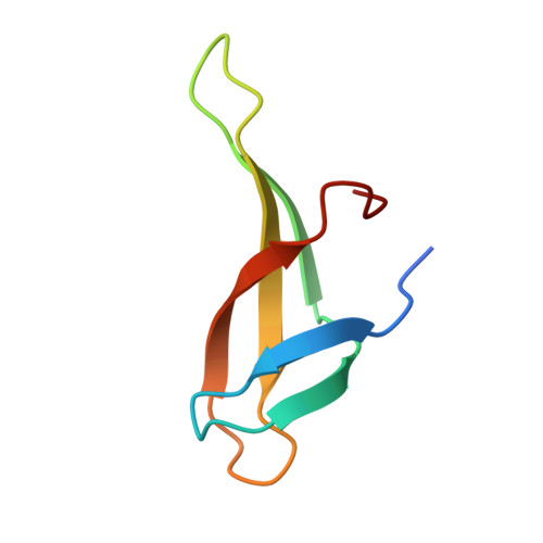
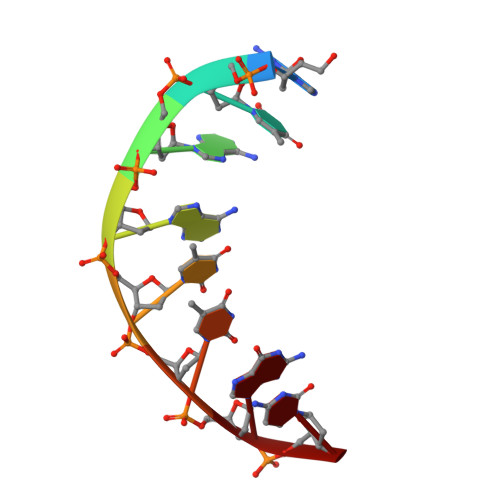
Sequence-Dependent T:G Base Pair Opening in DNA Double Helix Bound by Cren7, a Chromatin Protein Conserved among Crenarchaea

Tian, L.Zhang, Z.F.Wang, H.Zhao, M.Dong, Y.Gong, Y.

(2016) PLoS One 11: e0163361-e0163361

  • DOI: https://doi.org/10.1371/journal.pone.0163361
  • Primary Citation Related Structures: 
    5K07, 5K17

  • PubMed Abstract: 

    T:G base pair arising from spontaneous deamination of 5mC or polymerase errors is a great challenge for DNA repair of hyperthermophilic archaea, especially Crenarchaea. Most strains in this phylum lack the protein homologues responsible for the recognition of the mismatch in the DNA repair pathways. To investigate whether Cren7, a highly conserved chromatin protein in Crenarchaea, serves a role in the repair of T:G mispairs, the crystal structures of Cren7-GTAATTGC and Cren7-GTGATCGC complexes were solved at 2.0 Å and 2.1 Å. In our structures, binding of Cren7 to the AT-rich DNA duplex (GTAATTGC) induces opening of T2:G15 but not T10:G7 base pair. By contrast, both T:G mispairs in the GC-rich DNA duplex (GTGATCGC) retain the classic wobble type. Structural analysis also showed DNA helical changes of GTAATTGC, especially in the steps around the open T:G base pair, as compared to GTGATCGC or the matched DNAs. Surface plasmon resonance assays revealed a 4-fold lower binding affinity of Cren7 for GTAATTGC than that for GTGATCGC, which was dominantly contributed by the decrease of association rate. These results suggested that binding of Cren7 to DNA leads to T:G mispair opening in a sequence dependent manner, and therefore propose the potential roles of Cren7 in DNA repair.


  • Organizational Affiliation
    • Department of general surgery, Navy General Hospital, Beijing 100048, China.

Macromolecule Content 

  • Total Structure Weight: 11.56 kDa 
  • Atom Count: 872 
  • Modeled Residue Count: 73 
  • Deposited Residue Count: 76 
  • Unique protein chains: 1
  • Unique nucleic acid chains: 1

Macromolecules


Find similar proteins by:|  3D Structure
Entity ID: 1
MoleculeChains  Sequence LengthOrganismDetailsImage
Chromatin protein Cren760Saccharolobus solfataricus P2Mutation(s): 0 
Gene Names: creN7
UniProt
Find proteins for Q97ZE3 (Saccharolobus solfataricus (strain ATCC 35092 / DSM 1617 / JCM 11322 / P2))
Explore Q97ZE3 
Go to UniProtKB:  Q97ZE3
Entity Groups
Sequence Clusters30% Identity50% Identity70% Identity90% Identity95% Identity100% Identity
UniProt GroupQ97ZE3
Sequence Annotations
Expand
Reference Sequence
Find similar nucleic acids by:  Sequence
Entity ID: 2
MoleculeChains LengthOrganismImage
DNA (5'-D(*GP*TP*AP*AP*TP*TP*GP*C)-3')
B, C
8synthetic construct
Sequence Annotations
Expand
Reference Sequence

Experimental Data & Validation

Experimental Data

  • Method: X-RAY DIFFRACTION
  • Resolution: 2.00 Å
  • R-Value Free:  0.230 (Depositor), 0.227 (DCC) 
  • R-Value Work:  0.198 (Depositor), 0.199 (DCC) 
  • R-Value Observed: 0.199 (Depositor) 
Space Group: C 2 2 21
Unit Cell:
Length ( Å )Angle ( ˚ )
a = 51.795α = 90
b = 52.867β = 90
c = 90.689γ = 90
Software Package:
Software NamePurpose
PHENIXrefinement
HKL-2000data reduction
HKL-2000data scaling
PHASERphasing

Structure Validation

View Full Validation Report


Entry History 

Deposition Data

Revision History  (Full details and data files)

  • Version 1.0: 2017-05-24
    Type: Initial release
  • Version 1.1: 2023-11-08
    Changes: Data collection, Database references, Refinement description