5JXL

Cryo-EM structure of the flagellar hook of Campylobacter jejuni


Experimental Data Snapshot

  • Method: ELECTRON MICROSCOPY
  • Resolution: 3.50 Å
  • Aggregation State: FILAMENT 
  • Reconstruction Method: HELICAL 

wwPDB Validation   3D Report Full Report


This is version 1.4 of the entry. See complete history


Literature

Complete structure of the bacterial flagellar hook reveals extensive set of stabilizing interactions

Matsunami, H.Barker, C.S.Yoon, Y.H.Wolf, M.Samatey, F.A.

(2016) Nat Commun 7: 13425-13425

  • DOI: https://doi.org/10.1038/ncomms13425
  • Primary Citation of Related Structures:  
    5JXL

  • PubMed Abstract: 

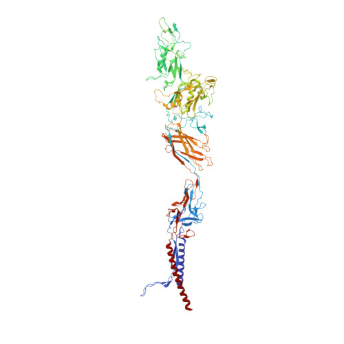
    The bacterial flagellar hook is a tubular helical structure made by the polymerization of multiple copies of a protein, FlgE. Here we report the structure of the hook from Campylobacter jejuni by cryo-electron microscopy at a resolution of 3.5 Å. On the basis of this structure, we show that the hook is stabilized by intricate inter-molecular interactions between FlgE molecules. Extra domains in FlgE, found only in Campylobacter and in related bacteria, bring more stability and robustness to the hook. Functional experiments suggest that Campylobacter requires an unusually strong hook to swim without its flagella being torn off. This structure reveals details of the quaternary organization of the hook that consists of 11 protofilaments. Previous study of the flagellar filament of Campylobacter by electron microscopy showed its quaternary structure made of seven protofilaments. Therefore, this study puts in evidence the difference between the quaternary structures of a bacterial filament and its hook.


  • Organizational Affiliation

    Trans-Membrane Trafficking Unit, Okinawa Institute of Science and Technology Graduate University, 1919-1 Tancha, Onna, Kunigami 904-0495, Japan.


Macromolecules
Find similar proteins by:  (by identity cutoff)  |  3D Structure
Entity ID: 1
MoleculeChains Sequence LengthOrganismDetailsImage
flagellar hook protein FlgE851Campylobacter jejuni subsp. jejuni 81116Mutation(s): 0 
UniProt
Find proteins for A0A1L1QK18 (Campylobacter jejuni subsp. jejuni serotype O:6 (strain 81116 / NCTC 11828))
Explore A0A1L1QK18 
Go to UniProtKB:  A0A1L1QK18
Entity Groups  
Sequence Clusters30% Identity50% Identity70% Identity90% Identity95% Identity100% Identity
UniProt GroupA0A1L1QK18
Sequence Annotations
Expand
  • Reference Sequence
Experimental Data & Validation

Experimental Data

  • Method: ELECTRON MICROSCOPY
  • Resolution: 3.50 Å
  • Aggregation State: FILAMENT 
  • Reconstruction Method: HELICAL 
EM Software:
TaskSoftware PackageVersion
RECONSTRUCTIONsegrefine3d0.83
MODEL REFINEMENTPHENIX1.9-1692

Structure Validation

View Full Validation Report


Entry History & Funding Information

Deposition Data


Funding OrganizationLocationGrant Number
Okinawa Institute of Science and Technology Graduate UniversityJapan--

Revision History  (Full details and data files)

  • Version 1.0: 2016-11-16
    Type: Initial release
  • Version 1.1: 2016-12-07
    Changes: Other
  • Version 1.2: 2018-07-25
    Changes: Data collection
  • Version 1.3: 2019-12-11
    Changes: Experimental preparation
  • Version 1.4: 2024-03-20
    Changes: Data collection, Database references, Derived calculations, Refinement description