5JUD

Crystal structure of glucosyl-3-phosphoglycerate synthase from Mycobacterium tuberculosis in complex with uridine-diphosphate (UDP) - GpgS*UDP


Experimental Data Snapshot

  • Method: X-RAY DIFFRACTION
  • Resolution: 2.59 Å
  • R-Value Free: 0.216 
  • R-Value Work: 0.195 
  • R-Value Observed: 0.196 

wwPDB Validation   3D Report Full Report


Ligand Structure Quality Assessment 


This is version 1.2 of the entry. See complete history


Literature

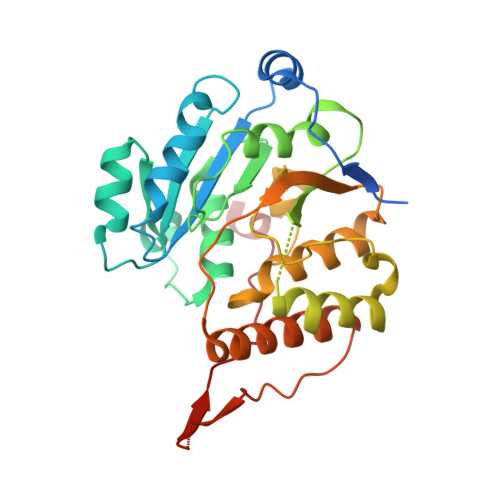
Structural Snapshots and Loop Dynamics along the Catalytic Cycle of Glycosyltransferase GpgS.

Albesa-Jove, D.Romero-Garcia, J.Sancho-Vaello, E.Contreras, F.X.Rodrigo-Unzueta, A.Comino, N.Carreras-Gonzalez, A.Arrasate, P.Urresti, S.Biarnes, X.Planas, A.Guerin, M.E.

(2017) Structure 25: 1034-1044.e3

  • DOI: https://doi.org/10.1016/j.str.2017.05.009
  • Primary Citation of Related Structures:  
    5JQX, 5JSX, 5JT0, 5JUC, 5JUD

  • PubMed Abstract: 

    Glycosyltransferases (GTs) play a central role in nature. They catalyze the transfer of a sugar moiety to a broad range of acceptor substrates. GTs are highly selective enzymes, allowing the recognition of subtle structural differences in the sequences and stereochemistry of their sugar and acceptor substrates. We report here a series of structural snapshots of the reaction center of the retaining glucosyl-3-phosphoglycerate synthase (GpgS). During this sequence of events, we visualize how the enzyme guides the substrates into the reaction center where the glycosyl transfer reaction takes place, and unveil the mechanism of product release, involving multiple conformational changes not only in the substrates/products but also in the enzyme. The structural data are further complemented by metadynamics free-energy calculations, revealing how the equilibrium of loop conformations is modulated along these itineraries. The information reported here represent an important contribution for the understanding of GT enzymes at the molecular level.


  • Organizational Affiliation

    Structural Biology Unit, CIC bioGUNE, Bizkaia Technology Park, Ed. 801A, 48160 Derio, Spain; Unidad de Biofísica, Centro Mixto Consejo Superior de Investigaciones Científicas - Universidad del País Vasco/Euskal Herriko Unibertsitatea (CSIC-UPV/EHU), Barrio Sarriena s/n, Leioa, Bizkaia 48940, Spain; Departamento de Bioquímica, Universidad del País Vasco, 48080 Bilbao, Spain; IKERBASQUE, Basque Foundation for Science, 48011 Bilbao, Spain.


Macromolecules
Find similar proteins by:  (by identity cutoff)  |  3D Structure
Entity ID: 1
MoleculeChains Sequence LengthOrganismDetailsImage
Glucosyl-3-phosphoglycerate synthase328Mycobacterium tuberculosis variant bovis AF2122/97Mutation(s): 0 
Gene Names: gpgSMb1240
EC: 2.4.1.266
UniProt
Find proteins for Q7U0E1 (Mycobacterium bovis (strain ATCC BAA-935 / AF2122/97))
Explore Q7U0E1 
Go to UniProtKB:  Q7U0E1
Entity Groups  
Sequence Clusters30% Identity50% Identity70% Identity90% Identity95% Identity100% Identity
UniProt GroupQ7U0E1
Sequence Annotations
Expand
  • Reference Sequence
Small Molecules
Ligands 1 Unique
IDChains Name / Formula / InChI Key2D Diagram3D Interactions
UDP
Query on UDP

Download Ideal Coordinates CCD File 
B [auth A]URIDINE-5'-DIPHOSPHATE
C9 H14 N2 O12 P2
XCCTYIAWTASOJW-XVFCMESISA-N
Experimental Data & Validation

Experimental Data

  • Method: X-RAY DIFFRACTION
  • Resolution: 2.59 Å
  • R-Value Free: 0.216 
  • R-Value Work: 0.195 
  • R-Value Observed: 0.196 
  • Space Group: I 41
Unit Cell:
Length ( Å )Angle ( ˚ )
a = 100.24α = 90
b = 100.24β = 90
c = 125.62γ = 90
Software Package:
Software NamePurpose
PHENIXrefinement
XDSdata reduction
XSCALEdata scaling
PHASERphasing

Structure Validation

View Full Validation Report


Ligand Structure Quality Assessment 


Entry History 

Deposition Data

Revision History  (Full details and data files)

  • Version 1.0: 2017-05-24
    Type: Initial release
  • Version 1.1: 2018-12-05
    Changes: Data collection, Database references
  • Version 1.2: 2024-01-10
    Changes: Data collection, Database references, Refinement description