5JQY | pdb_00005jqy

Aspartyl/Asparaginyl beta-hydroxylase (AspH)oxygenase and TPR domains in complex with manganese, N-oxalylglycine and factor X substrate peptide fragment(39mer-4Ser)


Experimental Data Snapshot

  • Method: X-RAY DIFFRACTION
  • Resolution: 1.99 Å
  • R-Value Free: 
    0.205 (Depositor), 0.208 (DCC) 
  • R-Value Work: 
    0.191 (Depositor), 0.195 (DCC) 
  • R-Value Observed: 
    0.192 (Depositor) 

Starting Model: experimental
View more details

wwPDB Validation 3D Report Full Report

Validation slider image for 5JQY

This is version 1.3 of the entry. See complete history

Literature

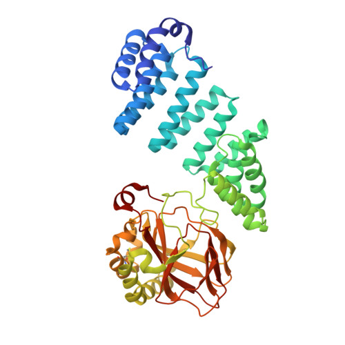
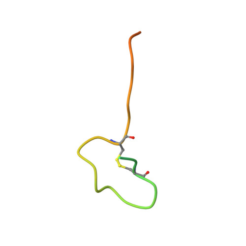
Aspartate/asparagine-beta-hydroxylase crystal structures reveal an unexpected epidermal growth factor-like domain substrate disulfide pattern.

Pfeffer, I.Brewitz, L.Krojer, T.Jensen, S.A.Kochan, G.T.Kershaw, N.J.Hewitson, K.S.McNeill, L.A.Kramer, H.Munzel, M.Hopkinson, R.J.Oppermann, U.Handford, P.A.McDonough, M.A.Schofield, C.J.

(2019) Nat Commun 10: 4910-4910

  • DOI: https://doi.org/10.1038/s41467-019-12711-7
  • Primary Citation Related Structures: 
    5APA, 5JQY, 5JZ6, 5JZ8, 5JZA, 5JZU, 6RK9

  • PubMed Abstract: 

    AspH is an endoplasmic reticulum (ER) membrane-anchored 2-oxoglutarate oxygenase whose C-terminal oxygenase and tetratricopeptide repeat (TPR) domains present in the ER lumen. AspH catalyses hydroxylation of asparaginyl- and aspartyl-residues in epidermal growth factor-like domains (EGFDs). Here we report crystal structures of human AspH, with and without substrate, that reveal substantial conformational changes of the oxygenase and TPR domains during substrate binding. Fe(II)-binding by AspH is unusual, employing only two Fe(II)-binding ligands (His679/His725). Most EGFD structures adopt an established fold with a conserved Cys1-3, 2-4, 5-6 disulfide bonding pattern; an unexpected Cys3-4 disulfide bonding pattern is observed in AspH-EGFD substrate complexes, the catalytic relevance of which is supported by studies involving stable cyclic peptide substrate analogues and by effects of Ca(II) ions on activity. The results have implications for EGFD disulfide pattern processing in the ER and will enable medicinal chemistry efforts targeting human 2OG oxygenases.


  • Organizational Affiliation
    • Chemistry Research Laboratory, University of Oxford, Mansfield Road, Oxford, OX1 3TA, UK.

Macromolecule Content 

  • Total Structure Weight: 53.86 kDa 
  • Atom Count: 3,878 
  • Modeled Residue Count: 447 
  • Deposited Residue Count: 468 
  • Unique protein chains: 2

Macromolecules

Find similar proteins by:|  3D Structure
Entity ID: 1
MoleculeChains  Sequence LengthOrganismDetailsImage
Aspartyl/asparaginyl beta-hydroxylase429Homo sapiensMutation(s): 0 
Gene Names: ASPHBAH
EC: 1.14.11.16
UniProt & NIH Common Fund Data Resources
Find proteins for Q12797 (Homo sapiens)
Explore Q12797 
Go to UniProtKB:  Q12797
PHAROS:  Q12797
GTEx:  ENSG00000198363 
Entity Groups
Sequence Clusters30% Identity50% Identity70% Identity90% Identity95% Identity100% Identity
UniProt GroupQ12797
Sequence Annotations
Expand
Reference Sequence
Find similar proteins by:|  3D Structure
Entity ID: 2
MoleculeChains  Sequence LengthOrganismDetailsImage
Coagulation factor X39Homo sapiensMutation(s): 4 
EC: 3.4.21.6
UniProt & NIH Common Fund Data Resources
Find proteins for P00742 (Homo sapiens)
Explore P00742 
Go to UniProtKB:  P00742
PHAROS:  P00742
GTEx:  ENSG00000126218 
Entity Groups
Sequence Clusters30% Identity50% Identity70% Identity90% Identity95% Identity100% Identity
UniProt GroupP00742
Sequence Annotations
Expand
Reference Sequence

Experimental Data & Validation

Experimental Data

  • Method: X-RAY DIFFRACTION
  • Resolution: 1.99 Å
  • R-Value Free:  0.205 (Depositor), 0.208 (DCC) 
  • R-Value Work:  0.191 (Depositor), 0.195 (DCC) 
  • R-Value Observed: 0.192 (Depositor) 
Space Group: P 21 21 21
Unit Cell:
Length ( Å )Angle ( ˚ )
a = 50.17α = 90
b = 89.54β = 90
c = 123.55γ = 90
Software Package:
Software NamePurpose
PHENIXrefinement
xia2data scaling
PDB_EXTRACTdata extraction
xia2data reduction
PHASERphasing

Structure Validation

View Full Validation Report


Entry History 

Deposition Data

Revision History  (Full details and data files)

  • Version 1.0: 2017-05-24
    Type: Initial release
  • Version 1.1: 2019-11-06
    Changes: Data collection, Database references
  • Version 1.2: 2024-01-10
    Changes: Data collection, Database references, Refinement description
  • Version 1.3: 2024-11-20
    Changes: Structure summary