


Crystal structure of Peroxiredoxin-1 from Schistosoma japonicum
Li, P., Li, Y.To be published.
Experimental Data Snapshot
Starting Model: experimental
View more details
wwPDB Validation 3D Report Full Report

Entity ID: 1 | |||||
|---|---|---|---|---|---|
| Molecule | Chains | Sequence Length | Organism | Details | Image |
| Thioredoxin peroxidase-1 | 190 | Schistosoma japonicum | Mutation(s): 0 Gene Names: SjTPx-1 EC: 1.11.1.24 | 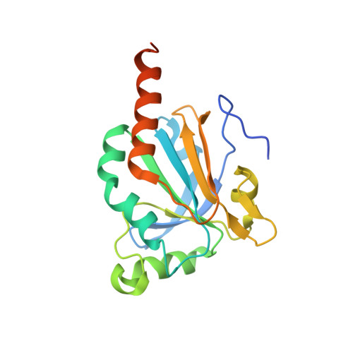 | |
UniProt | |||||
Find proteins for Q75UG3 (Schistosoma japonicum) Explore Q75UG3 Go to UniProtKB: Q75UG3 | |||||
Entity Groups | |||||
| Sequence Clusters | 30% Identity50% Identity70% Identity90% Identity95% Identity100% Identity | ||||
| UniProt Group | Q75UG3 | ||||
Sequence AnnotationsExpand | |||||
| |||||
| Length ( Å ) | Angle ( ˚ ) |
|---|---|
| a = 135.734 | α = 90 |
| b = 199.019 | β = 90 |
| c = 70.775 | γ = 90 |
| Software Name | Purpose |
|---|---|
| HKL-2000 | data scaling |
| PHENIX | refinement |
| PDB_EXTRACT | data extraction |
| HKL-2000 | data reduction |
| PHENIX | phasing |
| Funding Organization | Location | Grant Number |
|---|---|---|
| National Science Foundation of China | China | 31260206 |


