Cryoannealing-induced space-group transition of crystals of the carbonic anhydrase psCA3.
Pinard, M.A., Kurian, J.J., Aggarwal, M., Agbandje-McKenna, M., McKenna, R.(2016) Acta Crystallogr F Struct Biol Commun 72: 573-577
- PubMed: 27380376 Search on PubMedSearch on PubMed Central
- DOI: https://doi.org/10.1107/S2053230X16009286
- Primary Citation Related Structures:
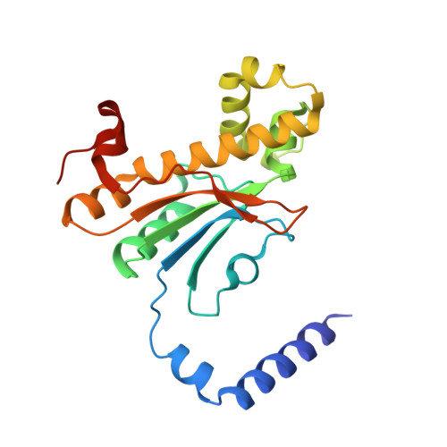
5JJ8 - PubMed Abstract:
Cryoannealing has been demonstrated to improve the diffraction quality and resolution of crystals of the β-carbonic anhydrase psCA3 concomitant with a change in space group. After initial flash-cooling in a liquid-nitrogen cryostream an X-ray diffraction data set from a psCA3 crystal was indexed in space group P21212 and was scaled to 2.6 Å resolution, but subsequent cryoannealing studies revealed induced protein rearrangements in the crystal contacts, which transformed the space group to I222, with a corresponding improvement of 0.7 Å in resolution. Although the change in diffraction resolution was significant, only minor changes in the psCA3 structure, which retained its catalytic `open' conformation, were observed. These findings demonstrate that cryoannealing can be successfully utilized to induce higher diffraction-quality crystals while maintaining enzymatically relevant conformations and may be useful as an experimental tool for structural studies of other enzymes where the initial diffraction quality is poor.
- Department of Biochemistry and Molecular Biology, University of Florida College of Medicine, 1200 Newell Drive, PO Box 100245, Gainesville, FL 32610, USA.
Organizational Affiliation: