5JGS | pdb_00005jgs

Human carbonic anhydrase II (F131Y/L198A) complexed with benzo[d]thiazole-2-sulfonamide


Experimental Data Snapshot

  • Method: X-RAY DIFFRACTION
  • Resolution: 1.11 Å
  • R-Value Free: 
    0.131 (Depositor), 0.135 (DCC) 
  • R-Value Work: 
    0.119 (Depositor), 0.121 (DCC) 
  • R-Value Observed: 
    0.119 (Depositor) 

Starting Model: experimental
View more details

wwPDB Validation 3D Report Full Report

Validation slider image for 5JGS

Ligand Structure Quality Assessment 


This is version 1.3 of the entry. See complete history

Literature

Water-Restructuring Mutations Can Reverse the Thermodynamic Signature of Ligand Binding to Human Carbonic Anhydrase.

Fox, J.M.Kang, K.Sastry, M.Sherman, W.Sankaran, B.Zwart, P.H.Whitesides, G.M.

(2017) Angew Chem Int Ed Engl 56: 3833-3837

  • DOI: https://doi.org/10.1002/anie.201609409
  • Primary Citation Related Structures: 
    5JDV, 5JE7, 5JEG, 5JEH, 5JEP, 5JES, 5JG3, 5JG5, 5JGS, 5JGT

  • PubMed Abstract: 

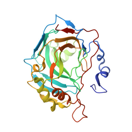
    This study uses mutants of human carbonic anhydrase (HCAII) to examine how changes in the organization of water within a binding pocket can alter the thermodynamics of protein-ligand association. Results from calorimetric, crystallographic, and theoretical analyses suggest that most mutations strengthen networks of water-mediated hydrogen bonds and reduce binding affinity by increasing the enthalpic cost and, to a lesser extent, the entropic benefit of rearranging those networks during binding. The organization of water within a binding pocket can thus determine whether the hydrophobic interactions in which it engages are enthalpy-driven or entropy-driven. Our findings highlight a possible asymmetry in protein-ligand association by suggesting that, within the confines of the binding pocket of HCAII, binding events associated with enthalpically favorable rearrangements of water are stronger than those associated with entropically favorable ones.


  • Organizational Affiliation
    • Department of Chemistry and Chemical Biology, Harvard University, 12 Oxford Street, Cambridge, MA, 02138, USA.

Macromolecule Content 

  • Total Structure Weight: 29.4 kDa 
  • Atom Count: 2,625 
  • Modeled Residue Count: 257 
  • Deposited Residue Count: 257 
  • Unique protein chains: 1

Macromolecules

Find similar proteins by:|  3D Structure
Entity ID: 1
MoleculeChains  Sequence LengthOrganismDetailsImage
Carbonic anhydrase 2A [auth B]257Homo sapiensMutation(s): 2 
Gene Names: CA2
EC: 4.2.1.1 (PDB Primary Data), 4.2.1.69 (UniProt)
UniProt & NIH Common Fund Data Resources
Find proteins for P00918 (Homo sapiens)
Explore P00918 
Go to UniProtKB:  P00918
PHAROS:  P00918
GTEx:  ENSG00000104267 
Entity Groups
Sequence Clusters30% Identity50% Identity70% Identity90% Identity95% Identity100% Identity
UniProt GroupP00918
Sequence Annotations
Expand
Reference Sequence

Experimental Data & Validation

Experimental Data

  • Method: X-RAY DIFFRACTION
  • Resolution: 1.11 Å
  • R-Value Free:  0.131 (Depositor), 0.135 (DCC) 
  • R-Value Work:  0.119 (Depositor), 0.121 (DCC) 
  • R-Value Observed: 0.119 (Depositor) 
Space Group: P 1 21 1
Unit Cell:
Length ( Å )Angle ( ˚ )
a = 42.35α = 90
b = 41.88β = 104.63
c = 72.06γ = 90
Software Package:
Software NamePurpose
PHENIXrefinement
xia2data reduction
PHASERphasing
xia2data scaling

Structure Validation

View Full Validation Report


Ligand Structure Quality Assessment 


Entry History 

Deposition Data

Revision History  (Full details and data files)

  • Version 1.0: 2017-01-11
    Type: Initial release
  • Version 1.1: 2017-03-15
    Changes: Database references
  • Version 1.2: 2017-03-29
    Changes: Database references
  • Version 1.3: 2023-09-27
    Changes: Data collection, Database references, Refinement description