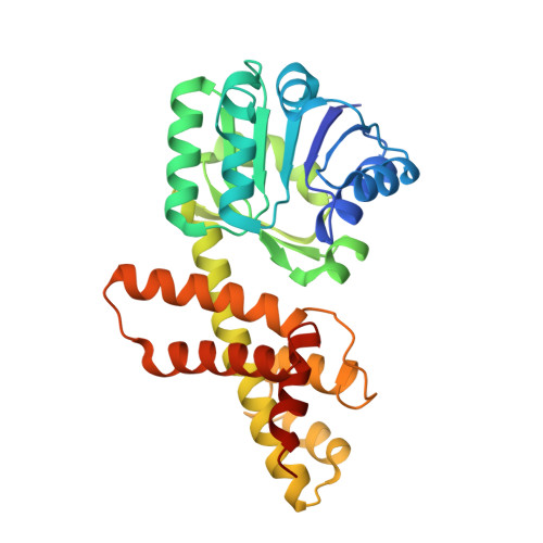
5JE8

The crystal structure of Bacillus cereus 3-hydroxyisobutyrate dehydrogenase in complex with NAD


Experimental Data Snapshot

  • Method: X-RAY DIFFRACTION
  • Resolution: 2.10 Å
  • R-Value Free: 0.243 
  • R-Value Work: 0.197 
  • R-Value Observed: 0.200 

wwPDB Validation   3D Report Full Report


Ligand Structure Quality Assessment 


This is version 1.2 of the entry. See complete history


Literature

Structural and biochemical characterization of the Bacillus cereus 3-hydroxyisobutyrate dehydrogenase

Park, S.C.Kim, P.H.Lee, G.S.Kang, S.G.Ko, H.J.Yoon, S.I.

(2016) Biochem Biophys Res Commun 474: 522-527

  • DOI: https://doi.org/10.1016/j.bbrc.2016.04.126
  • Primary Citation of Related Structures:  
    5JE8

  • PubMed Abstract: 

    The 3-hydroxyisobutyrate dehydrogenase (HIBADH) family catalyzes the NAD(+)- or NADP(+)-dependent oxidation of various β-hydroxyacid substrates into their cognate semialdehydes for diverse metabolic pathways. Because HIBADH group members exhibit different substrate specificities, the substrate-recognition mode of each enzyme should be individually characterized. In the current study, we report the biochemical and structural analysis of a HIBADH group enzyme from Bacillus cereus (bcHIBADH). bcHIBADH mediates a dehydrogenation reaction on S-3-hydroxyisobutyrate substrate with high catalytic efficiency in an NAD(+)-dependent manner; it also oxidizes l-serine and 3-hydroxypropionate with lower activity. bcHIBADH consists of two domains and is further assembled into a functional dimer rather than a tetramer that has been commonly observed in other prokaryotic HIBADH group members. In the bcHIBADH structure, the interdomain cleft forms a putative active site and simultaneously accommodates both an NAD(+) cofactor and a substrate mimic. Our structure-based comparative analysis highlights structural motifs that are important in the cofactor and substrate recognition of the HIBADH group.


  • Organizational Affiliation

    Division of Biomedical Convergence, College of Biomedical Science, Kangwon National University, Chuncheon 24341, Republic of Korea.


Macromolecules
Find similar proteins by:  (by identity cutoff)  |  3D Structure
Entity ID: 1
MoleculeChains Sequence LengthOrganismDetailsImage
3-hydroxyisobutyrate dehydrogenase
A, B, C, D
302Bacillus cereus ATCC 14579Mutation(s): 0 
Gene Names: BC_2289
EC: 1.1.1.31
UniProt
Find proteins for Q81DR6 (Bacillus cereus (strain ATCC 14579 / DSM 31 / CCUG 7414 / JCM 2152 / NBRC 15305 / NCIMB 9373 / NCTC 2599 / NRRL B-3711))
Explore Q81DR6 
Go to UniProtKB:  Q81DR6
Entity Groups  
Sequence Clusters30% Identity50% Identity70% Identity90% Identity95% Identity100% Identity
UniProt GroupQ81DR6
Sequence Annotations
Expand
  • Reference Sequence
Experimental Data & Validation

Experimental Data

  • Method: X-RAY DIFFRACTION
  • Resolution: 2.10 Å
  • R-Value Free: 0.243 
  • R-Value Work: 0.197 
  • R-Value Observed: 0.200 
  • Space Group: P 21 21 21
Unit Cell:
Length ( Å )Angle ( ˚ )
a = 46.444α = 90
b = 158.576β = 90
c = 163.138γ = 90
Software Package:
Software NamePurpose
REFMACrefinement
HKL-2000data reduction
HKL-2000data scaling
PHASERphasing

Structure Validation

View Full Validation Report


Ligand Structure Quality Assessment 


Entry History 

Deposition Data

Revision History  (Full details and data files)

  • Version 1.0: 2016-05-11
    Type: Initial release
  • Version 1.1: 2016-06-01
    Changes: Database references
  • Version 1.2: 2023-11-08
    Changes: Data collection, Database references, Derived calculations, Refinement description