Inhibition of Mycobacterium tuberculosis dihydrodipicolinate synthase by alpha-ketopimelic acid and its other structural analogues
Shrivastava, P., Navratna, V., Silla, Y., Dewangan, R.P., Pramanik, A., Chaudhary, S., Rayasam, G., Kumar, A., Gopal, B., Ramachandran, S.(2016) Sci Rep 6: 30827-30827
- PubMed: 27501775 Search on PubMedSearch on PubMed Central
- DOI: https://doi.org/10.1038/srep30827
- Primary Citation Related Structures:
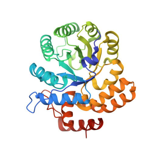
5J5D - PubMed Abstract:
The Mycobacterium tuberculosis dihydrodipicolinate synthase (Mtb-dapA) is an essential gene. Mtb-DapA catalyzes the aldol condensation between pyruvate and L-aspartate-beta-semialdehyde (ASA) to yield dihydrodipicolinate. In this work we tested the inhibitory effects of structural analogues of pyruvate on recombinant Mtb-DapA (Mtb-rDapA) using a coupled assay with recombinant dihydrodipicolinate reductase (Mtb-rDapB). Alpha-ketopimelic acid (α-KPA) showed maximum inhibition of 88% and IC50 of 21 μM in the presence of pyruvate (500 μM) and ASA (400 μM). Competition experiments with pyruvate and ASA revealed competition of α-KPA with pyruvate. Liquid chromatography-mass spectrometry (LC-MS) data with multiple reaction monitoring (MRM) showed that the relative abundance peak of final product, 2,3,4,5-tetrahydrodipicolinate, was decreased by 50%. Thermal shift assays showed 1 °C Tm shift of Mtb-rDapA upon binding α-KPA. The 2.4 Å crystal structure of Mtb-rDapA-α-KPA complex showed the interaction of critical residues at the active site with α-KPA. Molecular dynamics simulations over 500 ns of pyruvate docked to Mtb-DapA and of α-KPA-bound Mtb-rDapA revealed formation of hydrogen bonds with pyruvate throughout in contrast to α-KPA. Molecular descriptors analysis showed that ligands with polar surface area of 91.7 Å(2) are likely inhibitors. In summary, α-hydroxypimelic acid and other analogues could be explored further as inhibitors of Mtb-DapA.
- Functional Genomics Unit, Council of Scientific and Industrial Research-Institute of Genomics and Integrative Biology (CSIR-IGIB), South Campus, New Delhi 110025, India.
Organizational Affiliation: