Determination of crystal structures of proteins of unknown identity using a marathon molecular replacement procedure: structure of Stenotrophomonas maltophilia phosphate-binding protein.
Hatti, K., Gulati, A., Srinivasan, N., Murthy, M.R.(2016) Acta Crystallogr D Struct Biol 72: 1081-1089
- PubMed: 27710929 Search on PubMed
- DOI: https://doi.org/10.1107/S2059798316012419
- Primary Citation Related Structures:
5J1D - PubMed Abstract:
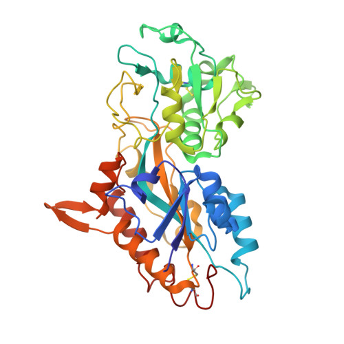
During the past decade, the authors have collected a few X-ray diffraction data sets from protein crystals that appeared to be easy cases of molecular replacement but failed to yield structures even after extensive trials. Here, the use of a large-scale molecular replacement method that explores all structurally characterized domains as phasing models to determine the structure corresponding to two data sets collected at 1.9 and 2.3 Å resolution is reported. These two structures were of the same protein independently crystallized in 2007 and 2011. The structures derived are virtually identical and were found to consist of two compact globular domains connected by a hinge. The high resolution of one of these data sets enabled inference of the amino-acid sequence from the electron-density map. The deduced sequence is nearly identical to that of a protein from the multidrug-resistant bacterium Stenotrophomonas maltophilia. Although the structure of this protein has not been determined previously, it is homologous to the well studied DING proteins which mediate the cellular uptake of phosphate ions. The final electron-density maps from both of the data sets revealed a large density at the interface of the two globular domains that is likely to represent a phosphate ion. Thus, the structure is likely to be that of a phosphate-binding protein encoded by the S. maltophilia genome (SmPBP; PDB entry 5j1d). The nature of the phosphate-binding site of SmPBP closely resembles that of Pseudomonas fluorescens DING (PfluDING), which displays remarkable discrimination between the closely similar phosphate and arsenate ions. The results presented here illustrate that routine crystallization trials may occasionally lead to the serendipitous crystallization of a protein of unknown identity and brute-force molecular replacement through `fold space' might allow the identification of the unknown protein.
- Molecular Biophysics Unit, Indian Institute of Science, Bengaluru, Karnataka 560 012, India.
Organizational Affiliation: