5J03

Crystal Structure of a chimeric Kv7.2 - Kv7.3 proximal C-terminal Domain in Complex with Calmodulin


Experimental Data Snapshot

  • Method: X-RAY DIFFRACTION
  • Resolution: 2.00 Å
  • R-Value Free: 0.228 
  • R-Value Work: 0.179 
  • R-Value Observed: 0.181 

wwPDB Validation   3D Report Full Report


This is version 2.0 of the entry. See complete history


Literature

Structural Insights into the M-Channel Proximal C-Terminus/Calmodulin Complex.

Strulovich, R.Tobelaim, W.S.Attali, B.Hirsch, J.A.

(2016) Biochemistry 55: 5353-5365

  • DOI: https://doi.org/10.1021/acs.biochem.6b00477
  • Primary Citation of Related Structures:  
    5J03

  • PubMed Abstract: 

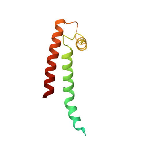
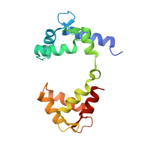
    The Kv7 (KCNQ) channel family, comprising voltage-gated potassium channels, plays major roles in fine-tuning cellular excitability by reducing firing frequency and controlling repolarization. Kv7 channels have a unique intracellular C-terminal (CT) domain bound constitutively by calmodulin (CaM). This domain plays key functions in channel tetramerization, trafficking, and gating. CaM binds to the proximal CT, comprising helices A and B. Kv7.2 and Kv7.3 are expressed in neural tissues. Together, they form the heterotetrameric M channel. We characterized Kv7.2, Kv7.3, and chimeric Kv7.3 helix A-Kv7.2 helix B (Q3A-Q2B) proximal CT/CaM complexes by solution methods at various Ca(2+)concentrations and determined them all to have a 1:1 stoichiometry. We then determined the crystal structure of the Q3A-Q2B/CaM complex at high Ca(2+) concentration to 2.0 Å resolution. CaM hugs the antiparallel coiled coil of helices A and B, braced together by an additional helix. The structure displays a hybrid apo-Ca(2+) CaM conformation even though four Ca(2+) ions are bound. Our results pinpoint unique interactions enabling the possible intersubunit pairing of Kv7.3 helix A and Kv7.2 helix B while underlining the potential importance of Kv7.3 helix A's role in stabilizing channel oligomerization. Also, the structure can be used to rationalize various channelopathic mutants. Functional testing of the chimeric channel found it to have a voltage-dependence similar to the M channel, thereby demonstrating helix A's importance in imparting gating properties.


  • Organizational Affiliation

    Department of Biochemistry and Molecular Biology, Institute of Structural Biology, George S. Wise Faculty of Life Sciences, ‡Department of Physiology and Pharmacology, Sackler Faculty of Medicine, and §Sagol School of Neuroscience, Tel Aviv University , Ramat Aviv 69978, Israel.


Macromolecules
Find similar proteins by:  (by identity cutoff)  |  3D Structure
Entity ID: 1
MoleculeChains Sequence LengthOrganismDetailsImage
Potassium voltage-gated channel subfamily KQT member 3,Potassium voltage-gated channel subfamily KQT member 2110Homo sapiensMutation(s): 0 
Gene Names: KCNQ3KCNQ2
UniProt & NIH Common Fund Data Resources
Find proteins for O43526 (Homo sapiens)
Explore O43526 
Go to UniProtKB:  O43526
PHAROS:  O43526
GTEx:  ENSG00000075043 
Find proteins for O43525 (Homo sapiens)
Explore O43525 
Go to UniProtKB:  O43525
PHAROS:  O43525
GTEx:  ENSG00000184156 
Entity Groups  
Sequence Clusters30% Identity50% Identity70% Identity90% Identity95% Identity100% Identity
UniProt GroupsO43526O43525
Sequence Annotations
Expand
  • Reference Sequence
Find similar proteins by:  (by identity cutoff)  |  3D Structure
Entity ID: 2
MoleculeChains Sequence LengthOrganismDetailsImage
Calmodulin149Homo sapiensMutation(s): 0 
Gene Names: 
CALM1CALMCAMCAM1CALM2CAM2CAMBCALM3CALML2CAM3...
CALM1CALMCAMCAM1CALM2CAM2CAMBCALM3CALML2CAM3CAMCCAMIII

UniProt & NIH Common Fund Data Resources
Find proteins for P0DP23 (Homo sapiens)
Explore P0DP23 
Go to UniProtKB:  P0DP23
PHAROS:  P0DP23
GTEx:  ENSG00000198668 
Entity Groups  
Sequence Clusters30% Identity50% Identity70% Identity90% Identity95% Identity100% Identity
UniProt GroupP0DP23
Sequence Annotations
Expand
  • Reference Sequence
Experimental Data & Validation

Experimental Data

  • Method: X-RAY DIFFRACTION
  • Resolution: 2.00 Å
  • R-Value Free: 0.228 
  • R-Value Work: 0.179 
  • R-Value Observed: 0.181 
  • Space Group: P 31 2 1
Unit Cell:
Length ( Å )Angle ( ˚ )
a = 47.665α = 90
b = 47.665β = 90
c = 182.216γ = 120
Software Package:
Software NamePurpose
PHENIXrefinement
XDSdata reduction
SCALAdata scaling
MOLREPphasing

Structure Validation

View Full Validation Report


Entry History & Funding Information

Deposition Data


Funding OrganizationLocationGrant Number
German Research FoundationGermany--
Israel Science FoundationIsrael1519/12

Revision History  (Full details and data files)

  • Version 1.0: 2016-09-07
    Type: Initial release
  • Version 1.1: 2016-10-05
    Changes: Database references
  • Version 2.0: 2019-10-16
    Changes: Atomic model, Author supporting evidence, Data collection