5IQY

Structure of apo-Dehydroascorbate Reductase from Pennisetum Glaucum phased by Iodide-SAD method


Experimental Data Snapshot

  • Method: X-RAY DIFFRACTION
  • Resolution: 2.40 Å
  • R-Value Free: 0.234 
  • R-Value Work: 0.184 
  • R-Value Observed: 0.188 

wwPDB Validation   3D Report Full Report


This is version 1.3 of the entry. See complete history


Literature

Non-native ligands define the active site of Pennisetum glaucum (L.) R. Br dehydroascorbate reductase

Das, B.K.Kumar, A.Maindola, P.Mahanty, S.Jain, S.K.Reddy, M.K.Arockiasamy, A.

(2016) Biochem Biophys Res Commun 473: 1152-1157

  • DOI: https://doi.org/10.1016/j.bbrc.2016.04.031
  • Primary Citation of Related Structures:  
    5EVO, 5IQY

  • PubMed Abstract: 

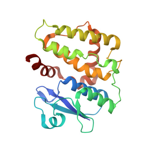
    Dehydroascorbate reductase (DHAR), a member of the glutathione-S-transferase (GST) family, reduces dehydroascorbate (DHA) to ascorbate (AsA; Vitamin-C) in a glutathione (GSH)-dependent manner and in doing so, replenishes the critical AsA pool of the cell. To understand the enzyme mechanism in detail, we determined the crystal structure of a plant DHAR from Pennisetum glaucum (PgDHAR) using Iodide-Single Anomalous Dispersion (SAD) and Molecular replacement methods, in two different space groups. Here, we show PgDHAR in complex with two non-native ligands, viz. an acetate bound at the G-site, which resembles the γ-carboxyl moiety of GSH, and a glycerol at the H-site, which shares the backbone of AsA. We also show that, in the absence of bound native substrates, these non-native ligands help define the critical 'hook points' in the DHAR enzyme active site. Further, our data suggest that these non-native ligands can act as the logical bootstrapping points for iterative design of inhibitors/analogs for DHARs.


  • Organizational Affiliation

    Membrane Protein Biology Group, ICGEB, Aruna Asaf Ali Marg, New Delhi, Delhi 110067, India; Department of Biotechnology, Jamia Hamdard University, New Delhi, Delhi, 110062, India.


Macromolecules
Find similar proteins by:  (by identity cutoff)  |  3D Structure
Entity ID: 1
MoleculeChains Sequence LengthOrganismDetailsImage
Dehydroascorbate reductase216Cenchrus americanusMutation(s): 0 
EC: 1.8.5.1
UniProt
Find proteins for U5XYA0 (Cenchrus americanus)
Explore U5XYA0 
Go to UniProtKB:  U5XYA0
Entity Groups  
Sequence Clusters30% Identity50% Identity70% Identity90% Identity95% Identity100% Identity
UniProt GroupU5XYA0
Sequence Annotations
Expand
  • Reference Sequence
Small Molecules
Ligands 1 Unique
IDChains Name / Formula / InChI Key2D Diagram3D Interactions
IOD
Query on IOD

Download Ideal Coordinates CCD File 
AA [auth A]
B [auth A]
C [auth A]
D [auth A]
E [auth A]
AA [auth A],
B [auth A],
C [auth A],
D [auth A],
E [auth A],
F [auth A],
G [auth A],
H [auth A],
I [auth A],
J [auth A],
K [auth A],
L [auth A],
M [auth A],
N [auth A],
O [auth A],
P [auth A],
Q [auth A],
R [auth A],
S [auth A],
T [auth A],
U [auth A],
V [auth A],
W [auth A],
X [auth A],
Y [auth A],
Z [auth A]
IODIDE ION
I
XMBWDFGMSWQBCA-UHFFFAOYSA-M
Experimental Data & Validation

Experimental Data

  • Method: X-RAY DIFFRACTION
  • Resolution: 2.40 Å
  • R-Value Free: 0.234 
  • R-Value Work: 0.184 
  • R-Value Observed: 0.188 
  • Space Group: C 2 2 21
Unit Cell:
Length ( Å )Angle ( ˚ )
a = 40.89α = 90
b = 132.08β = 90
c = 97.57γ = 90
Software Package:
Software NamePurpose
BUSTERrefinement
iMOSFLMdata reduction
Aimlessdata scaling
SHELXCDphasing

Structure Validation

View Full Validation Report


Entry History & Funding Information

Deposition Data


Funding OrganizationLocationGrant Number
DBTIndia--

Revision History  (Full details and data files)

  • Version 1.0: 2016-05-04
    Type: Initial release
  • Version 1.1: 2016-05-18
    Changes: Database references
  • Version 1.2: 2017-10-04
    Changes: Data collection, Database references, Derived calculations
  • Version 1.3: 2024-03-20
    Changes: Data collection, Database references