Structural Basis for Kinase-Mediated Macrolide Antibiotic Resistance.
Fong, D.H., Burk, D.L., Blanchet, J., Yan, A.Y., Berghuis, A.M.(2017) Structure 25: 750-761.e5
- PubMed: 28416110 Search on PubMed
- DOI: https://doi.org/10.1016/j.str.2017.03.007
- Primary Citation Related Structures:
5IGH, 5IGI, 5IGJ, 5IGP, 5IGR, 5IGS, 5IGT, 5IGU, 5IGV, 5IGW, 5IGY, 5IGZ, 5IH0, 5IH1, 5IWU - PubMed Abstract:
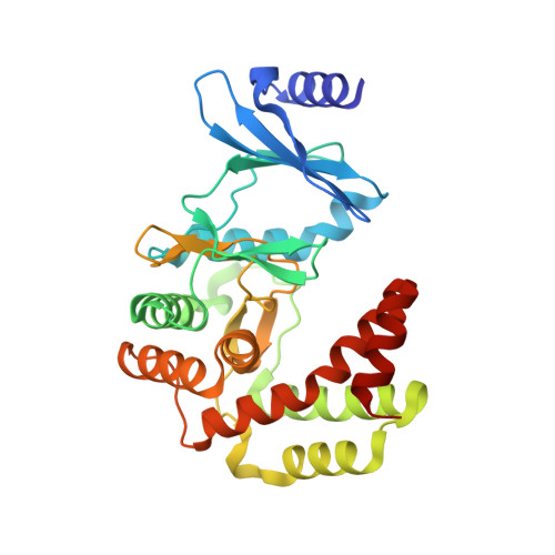
The macrolides are a class of antibiotic, characterized by a large macrocyclic lactone ring that can be inactivated by macrolide phosphotransferase enzymes. We present structures for MPH(2')-I and MPH(2')-II in the apo state, and in complex with GTP analogs and six different macrolides. These represent the first structures from the two main classes of macrolide phosphotransferases. The structures show that the enzymes are related to the aminoglycoside phosphotransferases, but are distinguished from them by the presence of a large interdomain linker that contributes to an expanded antibiotic binding pocket. This pocket is largely hydrophobic, with a negatively charged patch located at a conserved aspartate residue, rationalizing the broad-spectrum resistance conferred by the enzymes. Complementary mutation studies provide insights into factors governing substrate specificity. A comparison with macrolides bound to their natural target, the 50S ribosome, suggests avenues for next-generation antibiotic development.
- Department of Biochemistry, McGill University, Montréal, QC H3G 1Y6, Canada; Groupe de Recherche Axé sur la Structure des Protéines, McGill University, Montréal, QC H3G 0B1, Canada.
Organizational Affiliation: