5IFP | pdb_00005ifp

STRUCTURE OF BETA-GALACTOSIDASE FROM ASPERGILLUS NIGER


Experimental Data Snapshot

  • Method: X-RAY DIFFRACTION
  • Resolution: 1.71 Å
  • R-Value Free: 
    0.171 (Depositor), 0.181 (DCC) 
  • R-Value Work: 
    0.150 (Depositor), 0.162 (DCC) 
  • R-Value Observed: 
    0.151 (Depositor) 

Starting Model: experimental
View more details

wwPDB Validation 3D Report Full Report

Validation slider image for 5IFP

Ligand Structure Quality Assessment 


This is version 2.2 of the entry. See complete history

Literature

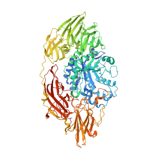
Structural features of Aspergillus niger beta-galactosidase define its activity against glycoside linkages.

Rico-Diaz, A.Ramirez-Escudero, M.Vizoso-Vazquez, A.Cerdan, M.E.Becerra, M.Sanz-Aparicio, J.

(2017) FEBS J 284: 1815-1829

  • DOI: https://doi.org/10.1111/febs.14083
  • Primary Citation Related Structures: 
    5IFP, 5IFT, 5IHR, 5JUV, 5MGC, 5MGD

  • PubMed Abstract: 

    β-Galactosidases are biotechnologically interesting enzymes that catalyze the hydrolysis or transgalactosylation of β-galactosides. Among them, the Aspergillus niger β-galactosidase (AnβGal) belongs to the glycoside hydrolase family 35 (GH35) and is widely used in the industry due to its high hydrolytic activity which is used to degrade lactose. We present here its three-dimensional structure in complex with different oligosaccharides, to illustrate the structural determinants of the broad specificity of the enzyme against different glycoside linkages. Remarkably, the residues Phe264, Tyr304, and Trp806 make a dynamic hydrophobic platform that accommodates the sugar at subsite +1 suggesting a main role on the recognition of structurally different substrates. Moreover, complexes with the trisaccharides show two potential subsites +2 depending on the substrate type. This feature and the peculiar shape of its wide cavity suggest that AnβGal might accommodate branched substrates from the complex net of polysaccharides composing the plant material in its natural environment. Relevant residues were selected and mutagenesis analyses were performed to evaluate their role in the catalytic performance and the hydrolase/transferase ratio of AnβGal. Thus, we generated mutants with improved transgalactosylation activity. In particular, the variant Y304F/Y355H/N357G/W806F displays a higher level of galacto-oligosaccharides production than the Aspergillus oryzae β-galactosidase, which is the preferred enzyme in the industry owing to its high transferase activity. Our results provide new knowledge on the determinants modulating specificity and the catalytic performance of fungal GH35 β-galactosidases. In turn, this fundamental background gives novel tools for the future improvement of these enzymes, which represent an interesting target for rational design. Structural data are available in PDB database under the accession numbers 5IFP (native form), 5IHR (in complex with 6GalGlu), 5IFT (in complex with 3GalGlu), 5JUV (in complex with 6GalGal), 5MGC (in complex with 4GalLac), and 5MGD (in complex with 6GalLac).


  • Organizational Affiliation
    • Grupo EXPRELA, Centro de Investigacións Científicas Avanzadas (CICA), Departamento de Bioloxía, Facultade de Ciencias, Universidade da Coruña, Spain.

Macromolecule Content 

  • Total Structure Weight: 117.15 kDa 
  • Atom Count: 8,920 
  • Modeled Residue Count: 968 
  • Deposited Residue Count: 1,013 
  • Unique protein chains: 1

Macromolecules

Find similar proteins by:|  3D Structure
Entity ID: 1
MoleculeChains  Sequence LengthOrganismDetailsImage
beta-galactosidase A1,013Aspergillus niger CBS 513.88Mutation(s): 0 
Gene Names: lacAAn01g12150
EC: 3.2.1.23
UniProt
Find proteins for A2QAN3 (Aspergillus niger (strain ATCC MYA-4892 / CBS 513.88 / FGSC A1513))
Explore A2QAN3 
Go to UniProtKB:  A2QAN3
Entity Groups
Sequence Clusters30% Identity50% Identity70% Identity90% Identity95% Identity100% Identity
UniProt GroupA2QAN3
Glycosylation
Glycosylation Sites: 10
Sequence Annotations
Expand
Reference Sequence

Oligosaccharides

Help  
Entity ID: 2
MoleculeChains Length2D Diagram GlycosylationD Interactions
alpha-D-mannopyranose-(1-2)-alpha-D-mannopyranose-(1-2)-alpha-D-mannopyranose-(1-3)-[alpha-D-mannopyranose-(1-3)-alpha-D-mannopyranose-(1-6)]beta-D-mannopyranose-(1-4)-2-acetamido-2-deoxy-beta-D-glucopyranose-(1-4)-2-acetamido-2-deoxy-beta-D-glucopyranose
B, C
8N-Glycosylation
Glycosylation Resources
GlyTouCan: G83582BK
GlyCosmos: G83582BK
GlyGen: G83582BK
Entity ID: 3
MoleculeChains Length2D Diagram GlycosylationD Interactions
2-acetamido-2-deoxy-beta-D-glucopyranose-(1-4)-2-acetamido-2-deoxy-beta-D-glucopyranose
D
2N-Glycosylation
Glycosylation Resources
GlyTouCan: G42666HT
GlyCosmos: G42666HT
GlyGen: G42666HT
Entity ID: 4
MoleculeChains Length2D Diagram GlycosylationD Interactions
alpha-D-mannopyranose-(1-2)-alpha-D-mannopyranose-(1-2)-alpha-D-mannopyranose-(1-3)-beta-D-mannopyranose-(1-4)-2-acetamido-2-deoxy-beta-D-glucopyranose-(1-4)-2-acetamido-2-deoxy-beta-D-glucopyranose
E
6N-Glycosylation
Glycosylation Resources
GlyTouCan: G01760ZU
GlyCosmos: G01760ZU
GlyGen: G01760ZU

Small Molecules

Ligands 5 Unique
IDChains Name / Formula / InChI Key2D Diagram3D Interactions
NAG

Query on NAG


Download:Ideal Coordinates CCD File
F [auth A]
G [auth A]
H [auth A]
I [auth A]
J [auth A]
F [auth A],
G [auth A],
H [auth A],
I [auth A],
J [auth A],
K [auth A]
2-acetamido-2-deoxy-beta-D-glucopyranose
C8 H15 N O6
OVRNDRQMDRJTHS-FMDGEEDCSA-N
BTB

Query on BTB


Download:Ideal Coordinates CCD File
O [auth A]2-[BIS-(2-HYDROXY-ETHYL)-AMINO]-2-HYDROXYMETHYL-PROPANE-1,3-DIOL
C8 H19 N O5
OWMVSZAMULFTJU-UHFFFAOYSA-N
SO4

Query on SO4


Download:Ideal Coordinates CCD File
L [auth A],
M [auth A]
SULFATE ION
O4 S
QAOWNCQODCNURD-UHFFFAOYSA-L
GOL

Query on GOL


Download:Ideal Coordinates CCD File
P [auth A],
Q [auth A],
R [auth A],
S [auth A],
T [auth A]
GLYCEROL
C3 H8 O3
PEDCQBHIVMGVHV-UHFFFAOYSA-N
CL

Query on CL


Download:Ideal Coordinates CCD File
N [auth A]CHLORIDE ION
Cl
VEXZGXHMUGYJMC-UHFFFAOYSA-M

Experimental Data & Validation

Experimental Data

  • Method: X-RAY DIFFRACTION
  • Resolution: 1.71 Å
  • R-Value Free:  0.171 (Depositor), 0.181 (DCC) 
  • R-Value Work:  0.150 (Depositor), 0.162 (DCC) 
  • R-Value Observed: 0.151 (Depositor) 
Space Group: P 21 21 21
Unit Cell:
Length ( Å )Angle ( ˚ )
a = 85.57α = 90
b = 111.421β = 90
c = 126.73γ = 90
Software Package:
Software NamePurpose
REFMACrefinement
MOSFLMdata reduction
Aimlessdata scaling
MOLREPphasing

Structure Validation

View Full Validation Report


Ligand Structure Quality Assessment 


Entry History 

Deposition Data

Revision History  (Full details and data files)

  • Version 1.0: 2017-04-19
    Type: Initial release
  • Version 1.1: 2017-06-28
    Changes: Database references
  • Version 2.0: 2020-07-29
    Type: Remediation
    Reason: Carbohydrate remediation
    Changes: Atomic model, Data collection, Derived calculations, Structure summary
  • Version 2.1: 2024-01-10
    Changes: Data collection, Database references, Derived calculations, Refinement description, Structure summary
  • Version 2.2: 2024-11-13
    Changes: Structure summary