5I79

Crystal structure of a beta-1,4-endoglucanase mutant from Aspergillus niger in complex with sugar


Experimental Data Snapshot

  • Method: X-RAY DIFFRACTION
  • Resolution: 2.35 Å
  • R-Value Free: 0.208 
  • R-Value Work: 0.147 
  • R-Value Observed: 0.150 

wwPDB Validation   3D Report Full Report


Ligand Structure Quality Assessment 


This is version 2.1 of the entry. See complete history


Literature

Functional and structural analysis of Pichia pastoris-expressed Aspergillus niger 1,4-beta-endoglucanase

Yan, J.Liu, W.Li, Y.Lai, H.L.Zheng, Y.Huang, J.W.Chen, C.C.Chen, Y.Jin, J.Li, H.Guo, R.T.

(2016) Biochem Biophys Res Commun 475: 8-12

  • DOI: https://doi.org/10.1016/j.bbrc.2016.05.012
  • Primary Citation of Related Structures:  
    5I77, 5I78, 5I79

  • PubMed Abstract: 

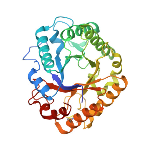
    Eukaryotic 1,4-β-endoglucanases (EC 3.2.1.4) have shown great potentials in many commercial applications because they effectively catalyze hydrolysis of cellulose, the main component of the plant cell wall. Here we expressed a glycoside hydrolase family (GH) 5 1,4-β-endoglucanase from Aspergillus niger (AnCel5A) in Pichia pastoris, which exhibits outstanding pH and heat stability. In order to further investigate the molecular mechanism of AnCel5A, apo-form and cellotetraose (CTT) complex enzyme crystal structures were solved to high resolution. AnCel5A folds into a typical (β/α)8-TIM barrel architecture, resembling other GH5 members. In the substrate binding cavity, CTT is found to bind to -4 - -1 subsites, and several polyethylene glycol molecules are found in positive subsites. In addition, several unique N-glycosylation motifs that may contribute to protein higher stability were observed from crystal structures. These results are of great importance for understanding the molecular mechanism of AnCel5A, and also provide guidance for further applications of the enzyme.


  • Organizational Affiliation

    School of Biotechnology, Jiangnan University, Wuxi 214122, China.


Macromolecules
Find similar proteins by:  (by identity cutoff)  |  3D Structure
Entity ID: 1
MoleculeChains Sequence LengthOrganismDetailsImage
Endo-beta-1, 4-glucanase
A, B
304Aspergillus nigerMutation(s): 1 
Gene Names: eg1
UniProt
Find proteins for O74706 (Aspergillus niger)
Explore O74706 
Go to UniProtKB:  O74706
Entity Groups  
Sequence Clusters30% Identity50% Identity70% Identity90% Identity95% Identity100% Identity
UniProt GroupO74706
Sequence Annotations
Expand
  • Reference Sequence
Oligosaccharides

Help

Entity ID: 2
MoleculeChains Length2D Diagram Glycosylation3D Interactions
beta-D-glucopyranose-(1-4)-beta-D-glucopyranose-(1-4)-beta-D-glucopyranose-(1-4)-beta-D-glucopyranose
C, D
4N/A
Glycosylation Resources
GlyTouCan:  G00025MO
GlyCosmos:  G00025MO
GlyGen:  G00025MO
Biologically Interesting Molecules (External Reference) 1 Unique
Experimental Data & Validation

Experimental Data

  • Method: X-RAY DIFFRACTION
  • Resolution: 2.35 Å
  • R-Value Free: 0.208 
  • R-Value Work: 0.147 
  • R-Value Observed: 0.150 
  • Space Group: C 1 2 1
Unit Cell:
Length ( Å )Angle ( ˚ )
a = 161.992α = 90
b = 51.332β = 95.2
c = 75.358γ = 90
Software Package:
Software NamePurpose
REFMACrefinement
HKL-2000data scaling
PHASERphasing
PDB_EXTRACTdata extraction
HKL-2000data reduction

Structure Validation

View Full Validation Report


Ligand Structure Quality Assessment 


Entry History 

Deposition Data

Revision History  (Full details and data files)

  • Version 1.0: 2016-12-21
    Type: Initial release
  • Version 2.0: 2020-07-29
    Type: Remediation
    Reason: Carbohydrate remediation
    Changes: Advisory, Atomic model, Data collection, Derived calculations, Non-polymer description, Structure summary
  • Version 2.1: 2023-11-08
    Changes: Data collection, Database references, Refinement description, Structure summary