5I74 | pdb_00005i74

X-ray structure of the ts3 human serotonin transporter complexed with Br-citalopram at the central site


Experimental Data Snapshot

  • Method: X-RAY DIFFRACTION
  • Resolution: 3.40 Å
  • R-Value Free: 
    0.296 (Depositor), 0.329 (DCC) 
  • R-Value Work: 
    0.249 (Depositor), 0.261 (DCC) 
  • R-Value Observed: 
    0.251 (Depositor) 

wwPDB Validation 3D Report Full Report

Validation slider image for 5I74

Ligand Structure Quality Assessment 


This is version 1.5 of the entry. See complete history

Literature

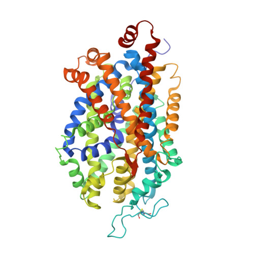
X-ray structures and mechanism of the human serotonin transporter.

Coleman, J.A.Green, E.M.Gouaux, E.

(2016) Nature 532: 334-339

  • DOI: https://doi.org/10.1038/nature17629
  • Primary Citation Related Structures: 
    5I66, 5I6X, 5I6Z, 5I71, 5I73, 5I74, 5I75

  • PubMed Abstract: 

    The serotonin transporter (SERT) terminates serotonergic signalling through the sodium- and chloride-dependent reuptake of neurotransmitter into presynaptic neurons. SERT is a target for antidepressant and psychostimulant drugs, which block reuptake and prolong neurotransmitter signalling. Here we report X-ray crystallographic structures of human SERT at 3.15 Å resolution bound to the antidepressants (S)-citalopram or paroxetine. Antidepressants lock SERT in an outward-open conformation by lodging in the central binding site, located between transmembrane helices 1, 3, 6, 8 and 10, directly blocking serotonin binding. We further identify the location of an allosteric site in the complex as residing at the periphery of the extracellular vestibule, interposed between extracellular loops 4 and 6 and transmembrane helices 1, 6, 10 and 11. Occupancy of the allosteric site sterically hinders ligand unbinding from the central site, providing an explanation for the action of (S)-citalopram as an allosteric ligand. These structures define the mechanism of antidepressant action in SERT, and provide blueprints for future drug design.


  • Organizational Affiliation
    • Vollum Institute, Oregon Health &Science University, Portland, Oregon 97239, USA.

Macromolecule Content 

  • Total Structure Weight: 110.57 kDa 
  • Atom Count: 7,610 
  • Modeled Residue Count: 974 
  • Deposited Residue Count: 984 
  • Unique protein chains: 3

Macromolecules

Find similar proteins by:|  3D Structure
Entity ID: 1
MoleculeChains  Sequence LengthOrganismDetailsImage
Sodium-dependent serotonin transporter549Homo sapiensMutation(s): 5 
Gene Names: SLC6A4HTTSERT
Membrane Entity: Yes 
UniProt & NIH Common Fund Data Resources
Find proteins for P31645 (Homo sapiens)
Explore P31645 
Go to UniProtKB:  P31645
PHAROS:  P31645
GTEx:  ENSG00000108576 
Entity Groups
Sequence Clusters30% Identity50% Identity70% Identity90% Identity95% Identity100% Identity
UniProt GroupP31645
Glycosylation
Glycosylation Sites: 2Go to GlyGen: P31645-1
Sequence Annotations
Expand
Reference Sequence
Find similar proteins by:|  3D Structure
Entity ID: 2
MoleculeChains  Sequence LengthOrganismDetailsImage
8B6 antibody, heavy chain221Mus musculusMutation(s): 0 
Find similar proteins by:|  3D Structure
Entity ID: 3
MoleculeChains  Sequence LengthOrganismDetailsImage
8B6 antibody, light chain214Mus musculusMutation(s): 0 

Small Molecules

Ligands 5 Unique
IDChains Name / Formula / InChI Key2D Diagram3D Interactions
CLR

Query on CLR


Download:Ideal Coordinates CCD File
F [auth A]CHOLESTEROL
C27 H46 O
HVYWMOMLDIMFJA-DPAQBDIFSA-N
69D

Query on 69D


Download:Ideal Coordinates CCD File
D [auth A](1S)-1-(4-bromophenyl)-1-[3-(dimethylamino)propyl]-1,3-dihydro-2-benzofuran-5-carbonitrile
C20 H21 Br N2 O
SOYXTZZNZQFLGF-FQEVSTJZSA-N
NAG

Query on NAG


Download:Ideal Coordinates CCD File
E [auth A],
I [auth A]
2-acetamido-2-deoxy-beta-D-glucopyranose
C8 H15 N O6
OVRNDRQMDRJTHS-FMDGEEDCSA-N
D12

Query on D12


Download:Ideal Coordinates CCD File
G [auth A]DODECANE
C12 H26
SNRUBQQJIBEYMU-UHFFFAOYSA-N
HEX

Query on HEX


Download:Ideal Coordinates CCD File
H [auth A]HEXANE
C6 H14
VLKZOEOYAKHREP-UHFFFAOYSA-N

Experimental Data & Validation

Experimental Data

  • Method: X-RAY DIFFRACTION
  • Resolution: 3.40 Å
  • R-Value Free:  0.296 (Depositor), 0.329 (DCC) 
  • R-Value Work:  0.249 (Depositor), 0.261 (DCC) 
  • R-Value Observed: 0.251 (Depositor) 
Space Group: C 2 2 21
Unit Cell:
Length ( Å )Angle ( ˚ )
a = 129.59α = 90
b = 164β = 90
c = 140.17γ = 90
Software Package:
Software NamePurpose
PHENIXrefinement
XDSdata reduction
XSCALEdata scaling
PHASERphasing

Structure Validation

View Full Validation Report


Ligand Structure Quality Assessment 


Entry History 

Deposition Data

Revision History  (Full details and data files)

  • Version 1.0: 2016-04-13
    Type: Initial release
  • Version 1.1: 2016-04-20
    Changes: Database references
  • Version 1.2: 2016-05-04
    Changes: Database references
  • Version 1.3: 2020-03-04
    Changes: Data collection, Database references, Derived calculations
  • Version 1.4: 2020-07-29
    Type: Remediation
    Reason: Carbohydrate remediation
    Changes: Data collection, Derived calculations, Structure summary
  • Version 1.5: 2024-11-06
    Changes: Data collection, Database references, Structure summary