5HYX

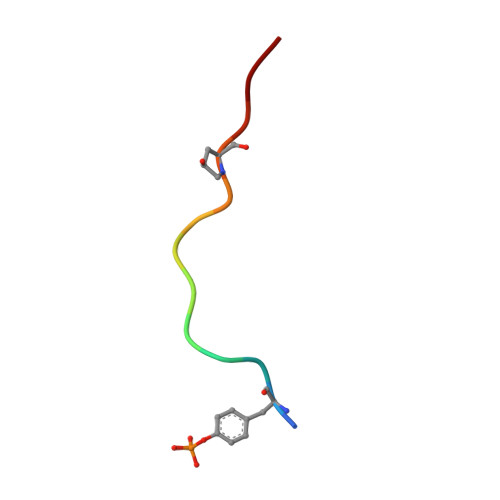
Plant peptide hormone receptor RGFR1 in complex with RGF1


Experimental Data Snapshot

  • Method: X-RAY DIFFRACTION
  • Resolution: 2.60 Å
  • R-Value Free: 0.233 
  • R-Value Work: 0.191 
  • R-Value Observed: 0.193 

wwPDB Validation   3D Report Full Report


Ligand Structure Quality Assessment 


This is version 2.0 of the entry. See complete history


Literature

Signature motif-guided identification of receptors for peptide hormones essential for root meristem growth

Song, W.Liu, L.Wang, J.Wu, Z.Zhang, H.Tang, J.Lin, G.Wang, Y.Wen, X.Li, W.Han, Z.Guo, H.Chai, J.

(2016) Cell Res 26: 674-685

  • DOI: https://doi.org/10.1038/cr.2016.62
  • Primary Citation of Related Structures:  
    5HYX

  • PubMed Abstract: 

    Peptide-mediated cell-to-cell signaling has crucial roles in coordination and definition of cellular functions in plants. Peptide-receptor matching is important for understanding the mechanisms underlying peptide-mediated signaling. Here we report the structure-guided identification of root meristem growth factor (RGF) receptors important for plant development. An assay based on a signature ligand recognition motif (Arg-x-Arg) conserved in a subfamily of leucine-rich repeat receptor kinases (LRR-RKs) identified the functionally uncharacterized LRR-RK At4g26540 as a receptor of RGF1 (RGFR1). We further solved the crystal structure of RGF1 in complex with the LRR domain of RGFR1 at a resolution of 2.6 Å, which reveals that the Arg-x-Gly-Gly (RxGG) motif is responsible for specific recognition of the sulfate group of RGF1 by RGFR1. Based on the RxGG motif, we identified additional four RGFRs. Participation of the five RGFRs in RGF-induced signaling is supported by biochemical and genetic data. We also offer evidence showing that SERKs function as co-receptors for RGFs. Taken together, our study identifies RGF receptors and co-receptors that can link RGF signals with their downstream components and provides a proof of principle for structure-based matching of LRR-RKs with their peptide ligands.


  • Organizational Affiliation

    Innovation Center for Structural Biology, Tsinghua-Peking Joint Center for Life Sciences, School of Life Sciences, Tsinghua University, Beijing 100084, China;


Macromolecules
Find similar proteins by:  (by identity cutoff)  |  3D Structure
Entity ID: 1
MoleculeChains Sequence LengthOrganismDetailsImage
Probable LRR receptor-like serine/threonine-protein kinase At4g26540A [auth B]633Arabidopsis thalianaMutation(s): 5 
Gene Names: At4g26540M3E9.30
EC: 2.7.11.1
UniProt
Find proteins for C0LGR3 (Arabidopsis thaliana)
Explore C0LGR3 
Go to UniProtKB:  C0LGR3
Entity Groups  
Sequence Clusters30% Identity50% Identity70% Identity90% Identity95% Identity100% Identity
UniProt GroupC0LGR3
Glycosylation
Glycosylation Sites: 3
Sequence Annotations
Expand
  • Reference Sequence

Find similar proteins by:  Sequence   |   3D Structure  

Entity ID: 2
MoleculeChains Sequence LengthOrganismDetailsImage
PTR-SER-ASN-PRO-GLY-HIS-HIS-PRO-HYP-ARG-HIS-ASNB [auth A]13ArabidopsisMutation(s): 0 
UniProt
Find proteins for Q3E880 (Arabidopsis thaliana)
Explore Q3E880 
Go to UniProtKB:  Q3E880
Entity Groups  
Sequence Clusters30% Identity50% Identity70% Identity90% Identity95% Identity100% Identity
UniProt GroupQ3E880
Sequence Annotations
Expand
  • Reference Sequence
Oligosaccharides

Help

Entity ID: 3
MoleculeChains Length2D Diagram Glycosylation3D Interactions
2-acetamido-2-deoxy-beta-D-glucopyranose-(1-4)-2-acetamido-2-deoxy-beta-D-glucopyranose
C
2N-Glycosylation
Glycosylation Resources
GlyTouCan:  G42666HT
GlyCosmos:  G42666HT
GlyGen:  G42666HT
Small Molecules
Ligands 1 Unique
IDChains Name / Formula / InChI Key2D Diagram3D Interactions
NAG
Query on NAG

Download Ideal Coordinates CCD File 
D [auth B],
E [auth B]
2-acetamido-2-deoxy-beta-D-glucopyranose
C8 H15 N O6
OVRNDRQMDRJTHS-FMDGEEDCSA-N
Modified Residues  2 Unique
IDChains TypeFormula2D DiagramParent
HZP
Query on HZP
B [auth A]L-PEPTIDE LINKINGC5 H9 N O3PRO
PTR
Query on PTR
B [auth A]L-PEPTIDE LINKINGC9 H12 N O6 PTYR
Experimental Data & Validation

Experimental Data

  • Method: X-RAY DIFFRACTION
  • Resolution: 2.60 Å
  • R-Value Free: 0.233 
  • R-Value Work: 0.191 
  • R-Value Observed: 0.193 
  • Space Group: H 3
Unit Cell:
Length ( Å )Angle ( ˚ )
a = 182.335α = 90
b = 182.335β = 90
c = 87.441γ = 120
Software Package:
Software NamePurpose
PHENIXrefinement
HKL-2000data scaling
PDB_EXTRACTdata extraction
HKL-2000data scaling
PHENIXphasing

Structure Validation

View Full Validation Report


Ligand Structure Quality Assessment 


Entry History 

Deposition Data

Revision History  (Full details and data files)

  • Version 1.0: 2017-01-11
    Type: Initial release
  • Version 2.0: 2020-07-29
    Type: Remediation
    Reason: Carbohydrate remediation
    Changes: Atomic model, Data collection, Derived calculations, Structure summary