5H03

Crystal structure of an ADP-ribosylating toxin BECa from C. perfringens


Experimental Data Snapshot

  • Method: X-RAY DIFFRACTION
  • Resolution: 1.89 Å
  • R-Value Free: 0.258 
  • R-Value Work: 0.209 
  • R-Value Observed: 0.212 

wwPDB Validation   3D Report Full Report


This is version 1.1 of the entry. See complete history


Literature

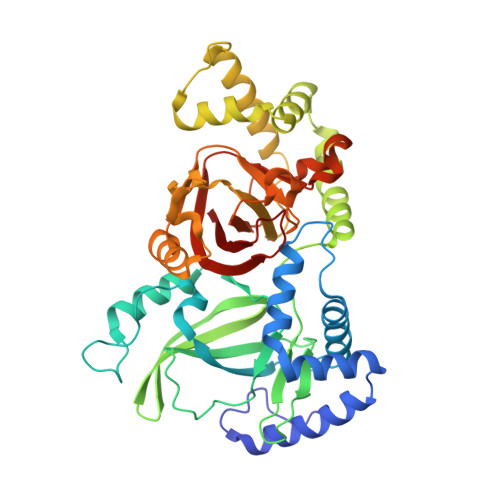
Crystal structure of the ADP-ribosylating component of BEC, the binary enterotoxin of Clostridium perfringens.

Kawahara, K.Yonogi, S.Munetomo, R.Oki, H.Yoshida, T.Kumeda, Y.Matsuda, S.Kodama, T.Ohkubo, T.Iida, T.Nakamura, S.

(2016) Biochem Biophys Res Commun 480: 261-267

  • DOI: https://doi.org/10.1016/j.bbrc.2016.10.042
  • Primary Citation of Related Structures:  
    5H03, 5H04

  • PubMed Abstract: 
  • Binary enterotoxin of Clostridium perfringens (BEC), consisting of the components BECa and BECb, was recently identified as a novel enterotoxin produced by C. perfringens that causes acute gastroenteritis in humans. Although the detailed mechanism of cell intoxication by BEC remains to be defined, BECa shows both NAD + -glycohydrolase and actin ADP-ribosyltransferase activities in the presence of NAD + ...

    Binary enterotoxin of Clostridium perfringens (BEC), consisting of the components BECa and BECb, was recently identified as a novel enterotoxin produced by C. perfringens that causes acute gastroenteritis in humans. Although the detailed mechanism of cell intoxication by BEC remains to be defined, BECa shows both NAD + -glycohydrolase and actin ADP-ribosyltransferase activities in the presence of NAD + . In this study, we determined the first crystal structure of BECa in its apo-state and in complex with NADH. The structure of BECa shows striking resemblance with other binary actin ADP-ribosylating toxins (ADPRTs), especially in terms of its overall protein fold and mechanisms of substrate recognition. We present a detailed picture of interactions between BECa and NADH, including bound water molecules located near the C1'-N glycosidic bond of NADH and the catalytically important ADP-ribosylating turn-turn (ARTT) loop. We observed that the conformational rearrangement of the ARTT loop, possibly triggered by a conformational change involving a conserved tyrosine residue coupled with substrate binding, plays a crucial role in catalysis by properly positioning a catalytic glutamate residue in the E-X-E motif of the ARTT loop in contact with the nucleophile. Our results for BECa provide insight into the common catalytic mechanism of the family of binary actin ADPRTs.


    Organizational Affiliation

    Research Institute for Microbial Diseases, Osaka University, Suita, Osaka, Japan. Electronic address: nshota@gen-info.osaka-u.ac.jp.


Macromolecules
Find similar proteins by:  (by identity cutoff)  |  3D Structure
Entity ID: 1
MoleculeChainsSequence LengthOrganismDetailsImage
Binary enterotoxin of Clostridium perfringens component aA419Clostridium perfringensMutation(s): 0 
Gene Names: becA
UniProt
Find proteins for X5I2D7 (Clostridium perfringens)
Explore X5I2D7 
Go to UniProtKB:  X5I2D7
Entity Groups  
Sequence Clusters30% Identity50% Identity70% Identity90% Identity95% Identity100% Identity
UniProt GroupX5I2D7
Protein Feature View
Expand
  • Reference Sequence
Experimental Data & Validation

Experimental Data

  • Method: X-RAY DIFFRACTION
  • Resolution: 1.89 Å
  • R-Value Free: 0.258 
  • R-Value Work: 0.209 
  • R-Value Observed: 0.212 
  • Space Group: I 2 2 2
Unit Cell:
Length ( Å )Angle ( ˚ )
a = 69.847α = 90
b = 94.002β = 90
c = 121.059γ = 90
Software Package:
Software NamePurpose
PHENIXrefinement
d*TREKdata scaling
PDB_EXTRACTdata extraction
CrystalCleardata reduction
PHASERphasing

Structure Validation

View Full Validation Report


Entry History 

Deposition Data

Revision History  (Full details and data files)

  • Version 1.0: 2016-11-02
    Type: Initial release
  • Version 1.1: 2016-11-09
    Changes: Database references