5GWW

Structure of MoeN5-Sso7d fusion protein in complex with a permethylated substrate analogue


Experimental Data Snapshot

  • Method: X-RAY DIFFRACTION
  • Resolution: 2.30 Å
  • R-Value Free: 0.221 
  • R-Value Work: 0.174 

wwPDB Validation   3D Report Full Report


Ligand Structure Quality Assessment 


This is version 1.2 of the entry. See complete history


Literature

Complex structures of MoeN5 with substrate analogues suggest sequential catalytic mechanism.

Zhang, L.Ko, T.-P.Malwal, S.R.Liu, W.Zhou, S.Yu, X.Oldfield, E.Guo, R.-T.Chen, C.-C.

(2019) Biochem Biophys Res Commun 511: 800-805

  • DOI: https://doi.org/10.1016/j.bbrc.2019.02.131
  • Primary Citation of Related Structures:  
    5GWV, 5GWW, 6J8V, 6J8W

  • PubMed Abstract: 

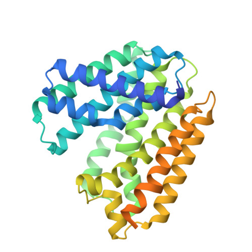
    The antibiotic moenomycin A is a phosphoglycerate derivative with a C 25 -moenocinyl chain and a branched oligosaccharide. Formation of the C 25 -chain is catalyzed by the enzyme MoeN5 with geranyl pyrophosphate (GPP) and the sugar-linked 2-Z,E-farnesyl-3-phosphoglycerate (FPG) as its substrates. Previous complex crystal structures with GPP and long-chain alkyl glycosides suggested that GPP binds to the S1 site in a similar way as in most other α-helical prenyltransferases (PTs), and FPG is likely to assume a bent conformation in the S2 site. However, two FPG derivatives synthesized in the current study were found in the S1 site rather than S2 in their complex crystal structures with MoeN5. Apparently S1 is the preferred site for prenyl-containing ligand, and S2 binding may proceed only after S1 is occupied. Thus, like most trans-type PTs, MoeN5 may employ a sequential ionization-condensation-elimination mechanism that involves a carbocation intermediate.


  • Organizational Affiliation

    University of Chinese Academy of Sciences, Beijing, 100049, China; Tianjin Institute of Industrial Biotechnology, Chinese Academy of Sciences, Tianjin, 300308, China.


Macromolecules
Find similar proteins by:  (by identity cutoff)  |  3D Structure
Entity ID: 1
MoleculeChains Sequence LengthOrganismDetailsImage
MoeN5,DNA-binding protein 7d
A, B, C, D
343Streptomyces viridosporusSaccharolobus solfataricus P2
This entity is chimeric
Mutation(s): 0 
Gene Names: moeN5sso7dsso7d-1SSO10610
UniProt
Find proteins for A0A010 (Streptomyces viridosporus)
Explore A0A010 
Go to UniProtKB:  A0A010
Find proteins for P39476 (Saccharolobus solfataricus (strain ATCC 35092 / DSM 1617 / JCM 11322 / P2))
Explore P39476 
Go to UniProtKB:  P39476
Entity Groups  
Sequence Clusters30% Identity50% Identity70% Identity90% Identity95% Identity100% Identity
UniProt GroupsA0A010P39476
Sequence Annotations
Expand
  • Reference Sequence
Small Molecules
Ligands 1 Unique
IDChains Name / Formula / InChI Key2D Diagram3D Interactions
7B5
Query on 7B5

Download Ideal Coordinates CCD File 
E [auth D]methyl (2R)-3-dimethoxyphosphoryloxy-2-[(2Z,6E)-3,7,11-trimethyldodeca-2,6,10-trienoxy]propanoate
C21 H37 O7 P
KGEZAZGNLOIEBB-KXPRUBRRSA-N
Experimental Data & Validation

Experimental Data

  • Method: X-RAY DIFFRACTION
  • Resolution: 2.30 Å
  • R-Value Free: 0.221 
  • R-Value Work: 0.174 
  • Space Group: C 2 2 2
Unit Cell:
Length ( Å )Angle ( ˚ )
a = 137.857α = 90
b = 217.331β = 90
c = 104.144γ = 90
Software Package:
Software NamePurpose
CNSrefinement
HKL-2000data reduction
HKL-2000data scaling
CNSphasing

Structure Validation

View Full Validation Report


Ligand Structure Quality Assessment 


Entry History & Funding Information

Deposition Data


Funding OrganizationLocationGrant Number
Ministry of Science and TechnologyTaiwanMOST105--0210-01-12-01

Revision History  (Full details and data files)

  • Version 1.0: 2017-09-20
    Type: Initial release
  • Version 1.1: 2019-04-03
    Changes: Data collection, Database references
  • Version 1.2: 2023-11-08
    Changes: Data collection, Database references, Refinement description