5GOD | pdb_00005god

Lys27-linked di-ubiquitin


Experimental Data Snapshot

  • Method: X-RAY DIFFRACTION
  • Resolution: 1.15 Å
  • R-Value Free: 
    0.222 (Depositor), 0.222 (DCC) 
  • R-Value Work: 
    0.202 (Depositor) 
  • R-Value Observed: 
    0.203 (Depositor) 

Starting Model: experimental
View more details

wwPDB Validation 3D Report Full Report

Validation slider image for 5GOD

This is version 1.4 of the entry. See complete history

Literature

Monomer/Oligomer Quasi-Racemic Protein Crystallography

Gao, S.Pan, M.Zheng, Y.Huang, Y.Zheng, Q.Sun, D.Lu, L.Tan, X.Tan, X.Lan, H.Wang, J.Wang, T.Wang, J.Liu, L.

(2016) J Am Chem Soc 138: 14497-14502

  • DOI: https://doi.org/10.1021/jacs.6b09545
  • Primary Citation Related Structures: 
    5GO7, 5GO8, 5GOB, 5GOC, 5GOD, 5GOG, 5GOH, 5GOI, 5GOJ, 5GOK

  • PubMed Abstract: 

    Racemic or quasi-racemic crystallography recently emerges as a useful technology for solution of the crystal structures of biomacromolecules. It remains unclear to what extent the biomacromolecules of opposite handedness can differ from each other in racemic or quasi-racemic crystallography. Here we report a finding that monomeric d-ubiquitin (Ub) has propensity to cocrystallize with different dimers, trimers, and even a tetramer of l-Ub. In these cocrystals the unconnected monomeric d-Ubs can self-assemble to form pseudomirror images of different oligomers of l-Ub. This monomer/oligomer cocrystallization phenomenon expands the concept of racemic crystallography. Using the monomer/oligomer cocrystallization technology we obtained, for the first time the X-ray structures of linear M1-linked tri- and tetra-Ubs and a K11/K63-branched tri-Ub.


  • Organizational Affiliation
    • Tsinghua-Peking Center for Life Sciences, Ministry of Education Key Laboratory of Bioorganic Phosphorus Chemistry and Chemical Biology, Department of Chemistry and ‡State Key Laboratory of Biomembrane and Membrane Biotechnology, Center for Structural Biology, School of Life Sciences, Tsinghua University , Beijing 100084, China.

Macromolecule Content 

  • Total Structure Weight: 34.28 kDa 
  • Atom Count: 2,745 
  • Modeled Residue Count: 304 
  • Deposited Residue Count: 304 
  • Unique protein chains: 2

Macromolecules

Find similar proteins by:|  3D Structure
Entity ID: 1
MoleculeChains  Sequence LengthOrganismDetailsImage
Ubiquitin
A, B
76Homo sapiensMutation(s): 0 
UniProt & NIH Common Fund Data Resources
Find proteins for P0CG47 (Homo sapiens)
Explore P0CG47 
Go to UniProtKB:  P0CG47
PHAROS:  P0CG47
GTEx:  ENSG00000170315 
Entity Groups
Sequence Clusters30% Identity50% Identity70% Identity90% Identity95% Identity100% Identity
UniProt GroupP0CG47
Sequence Annotations
Expand
Reference Sequence
Find similar proteins by:|  3D Structure
Entity ID: 2
MoleculeChains  Sequence LengthOrganismDetailsImage
D-ubiquitin
C, D
76Homo sapiensMutation(s): 0 
UniProt & NIH Common Fund Data Resources
Find proteins for P62987 (Homo sapiens)
Explore P62987 
Go to UniProtKB:  P62987
PHAROS:  P62987
GTEx:  ENSG00000221983 
Entity Groups
UniProt GroupP62987
Sequence Annotations
Expand
Reference Sequence

Small Molecules

Ligands 1 Unique
IDChains Name / Formula / InChI Key2D Diagram3D Interactions
MG

Query on MG


Download:Ideal Coordinates CCD File
E [auth B]MAGNESIUM ION
Mg
JLVVSXFLKOJNIY-UHFFFAOYSA-N
Modified Residues  17 Unique
IDChains TypeFormula2D DiagramParent
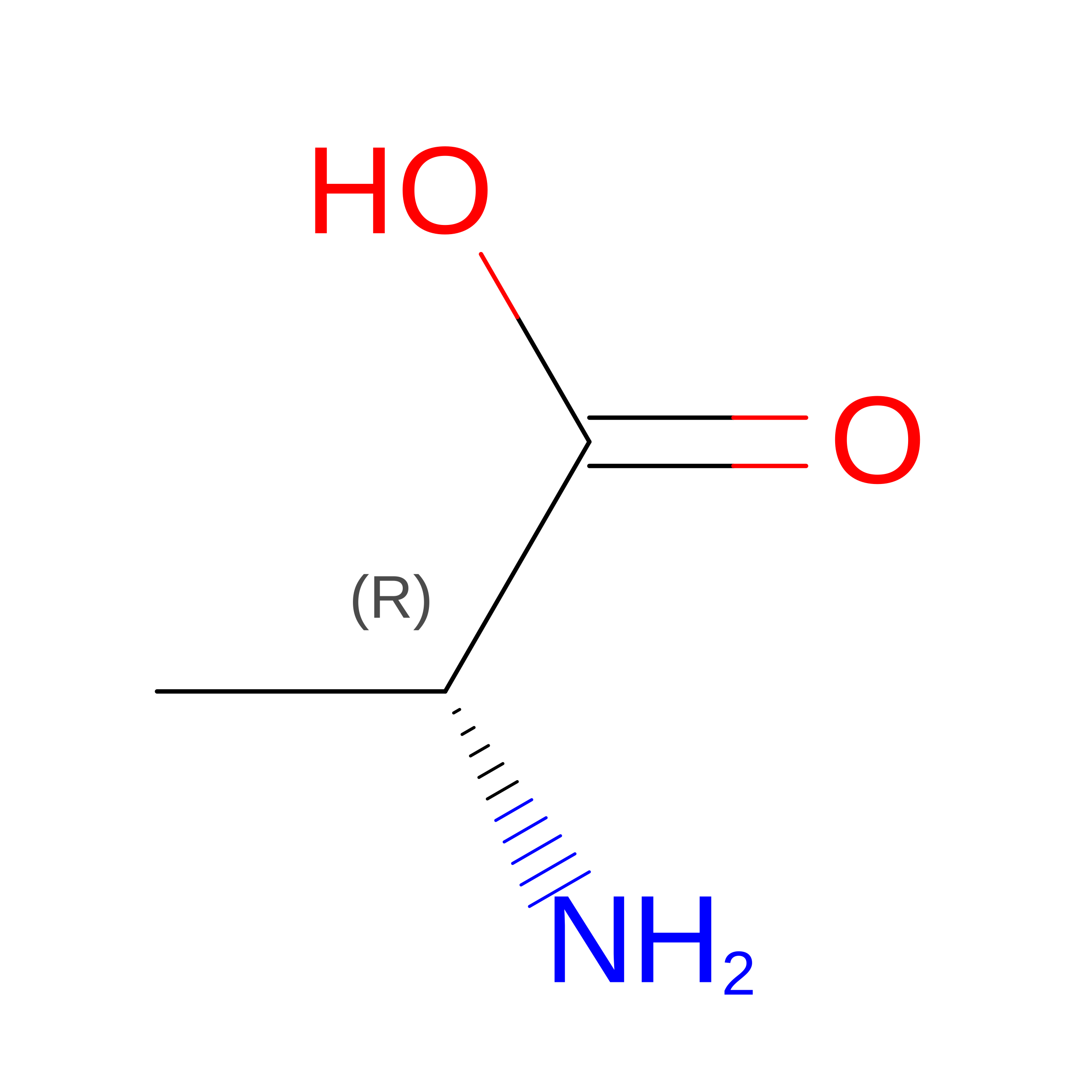
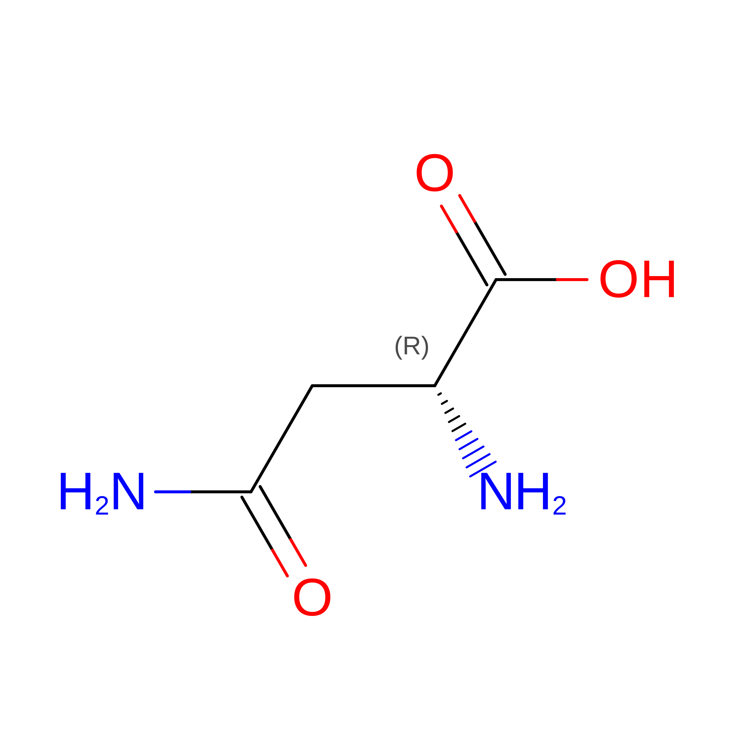
DAL
Query on DAL
C, D
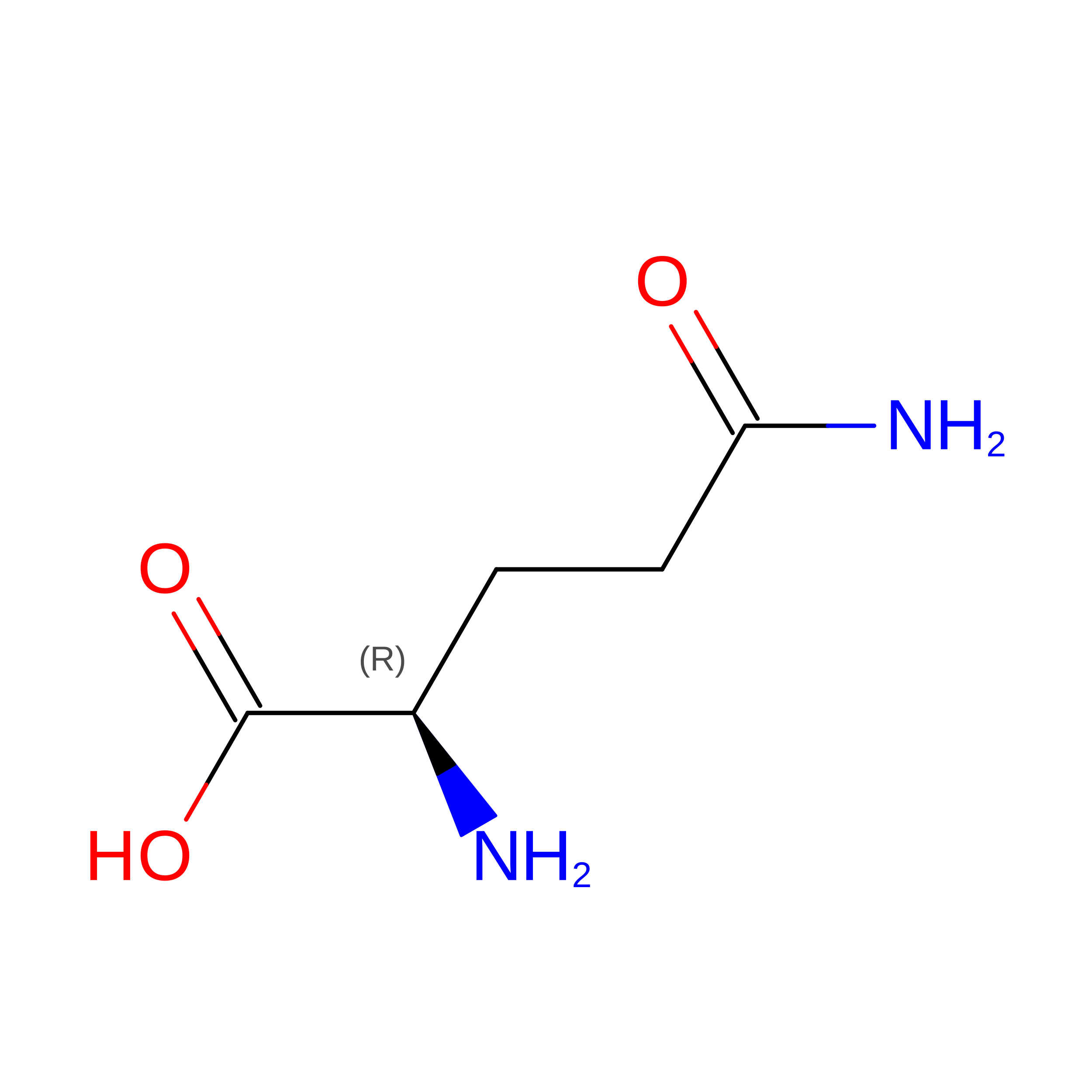
D-PEPTIDE LINKINGC3 H7 N O2Not Available
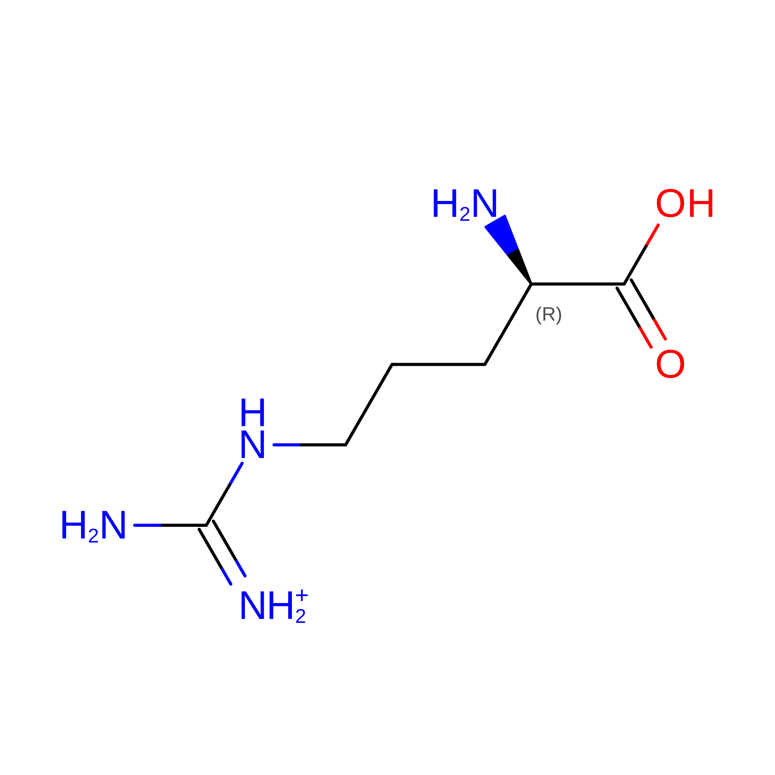
DAR
Query on DAR
C, D
D-PEPTIDE LINKINGC6 H15 N4 O2Not Available
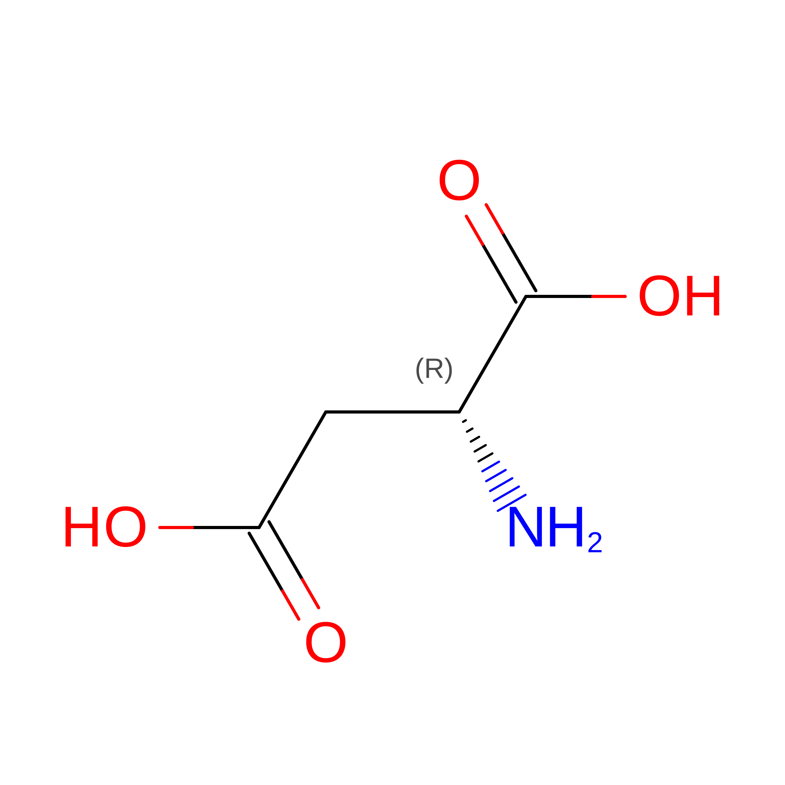
DAS
Query on DAS
C, D
D-PEPTIDE LINKINGC4 H7 N O4Not Available
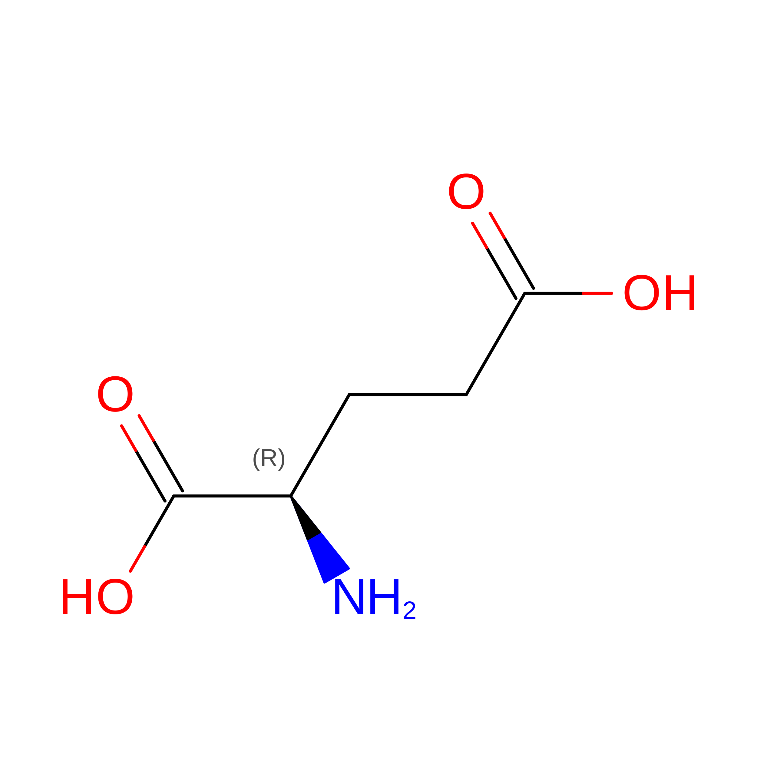
DGL
Query on DGL
C, D
D-PEPTIDE LINKINGC5 H9 N O4Not Available
DGN
Query on DGN
C, D
D-PEPTIDE LINKINGC5 H10 N2 O3Not Available
DHI
Query on DHI
C, D
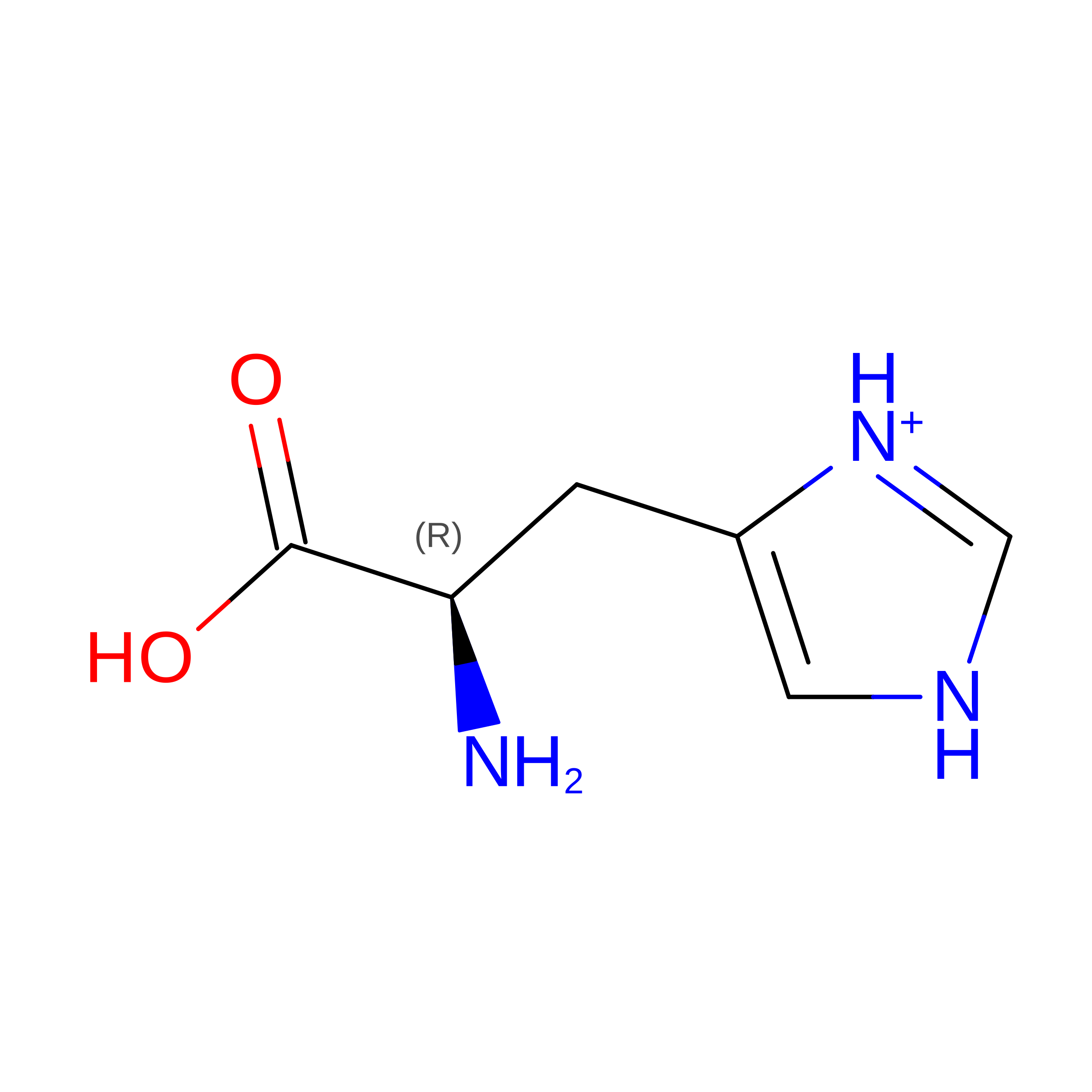
D-PEPTIDE LINKINGC6 H10 N3 O2Not Available
DIL
Query on DIL
C, D
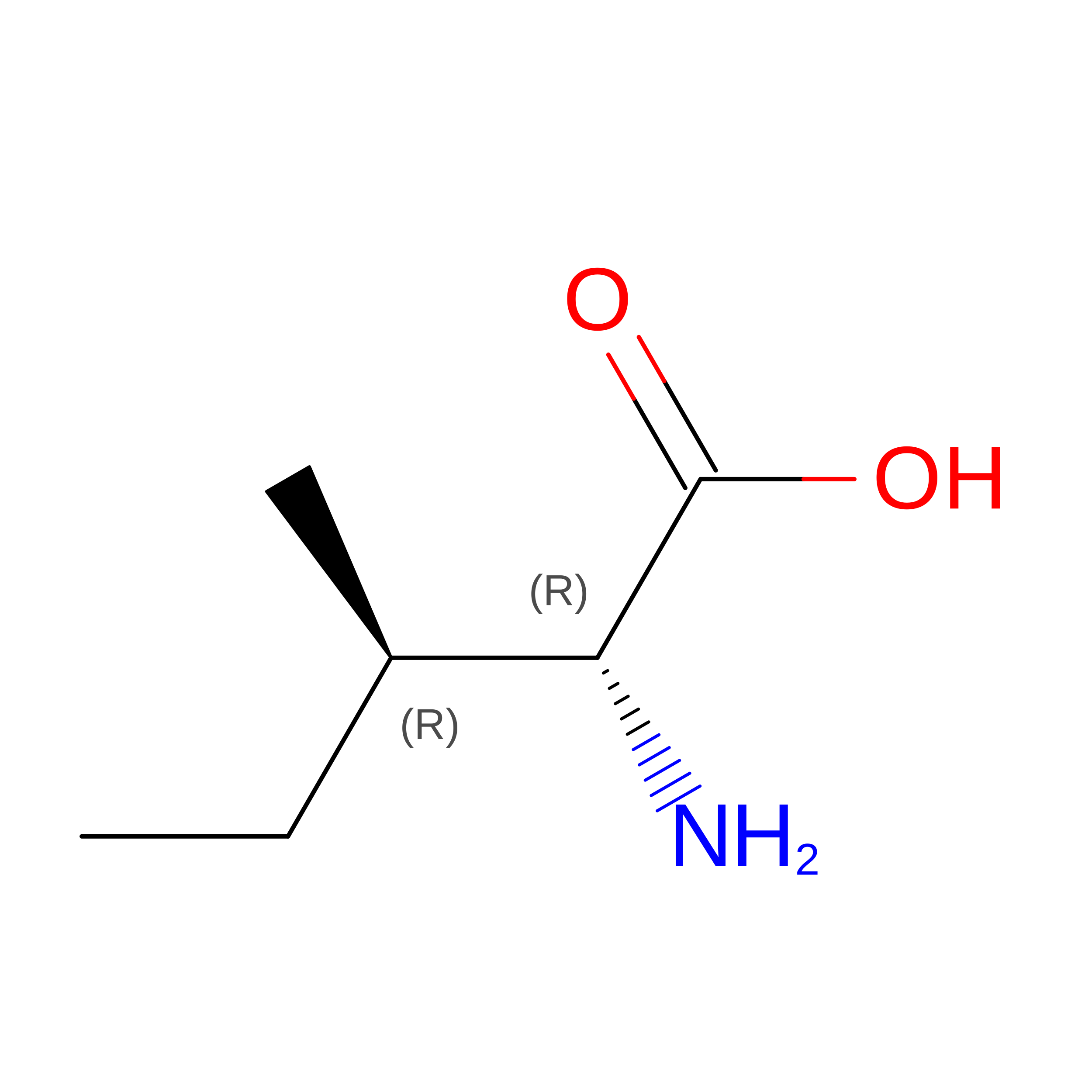
D-PEPTIDE LINKINGC6 H13 N O2Not Available
DLE
Query on DLE
C, D
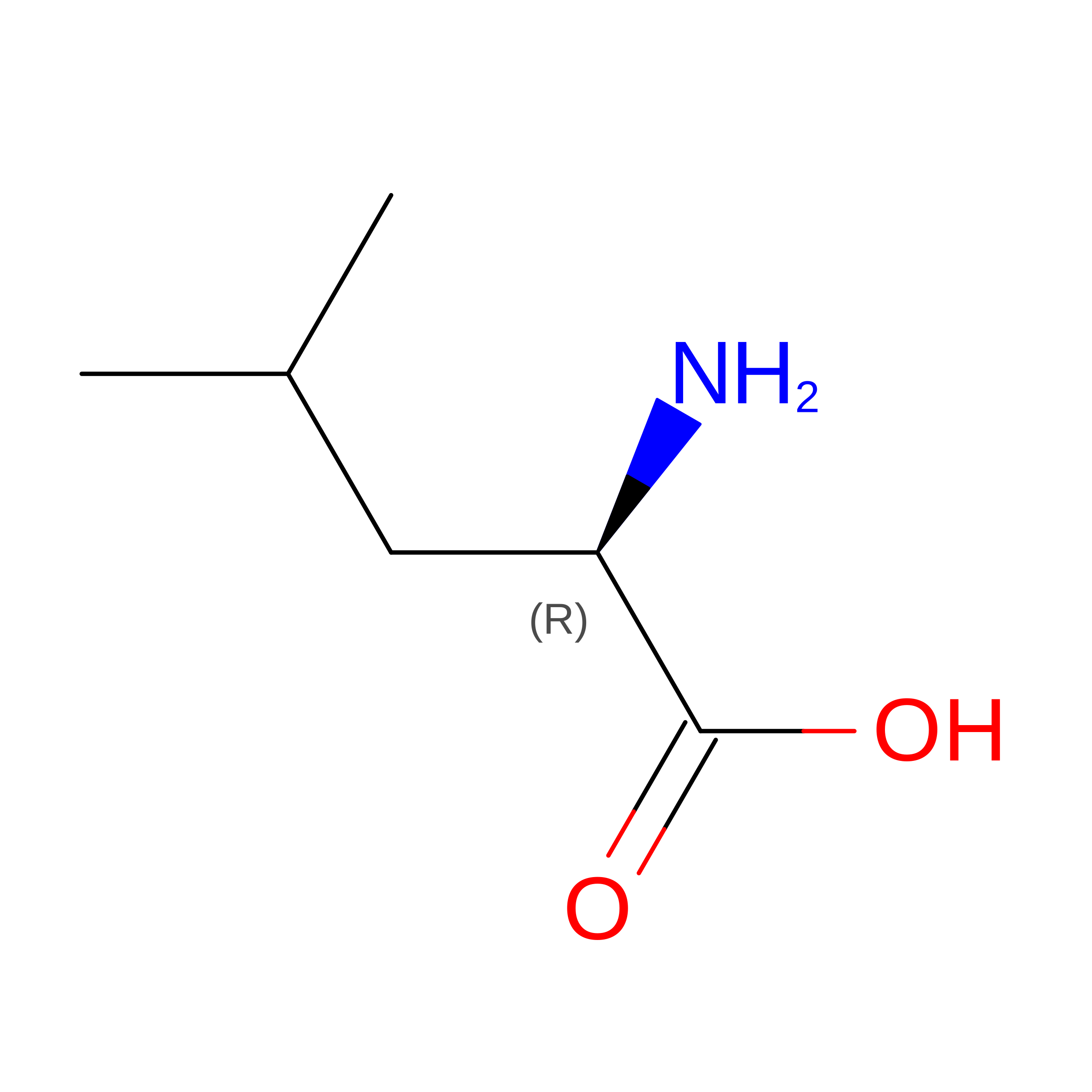
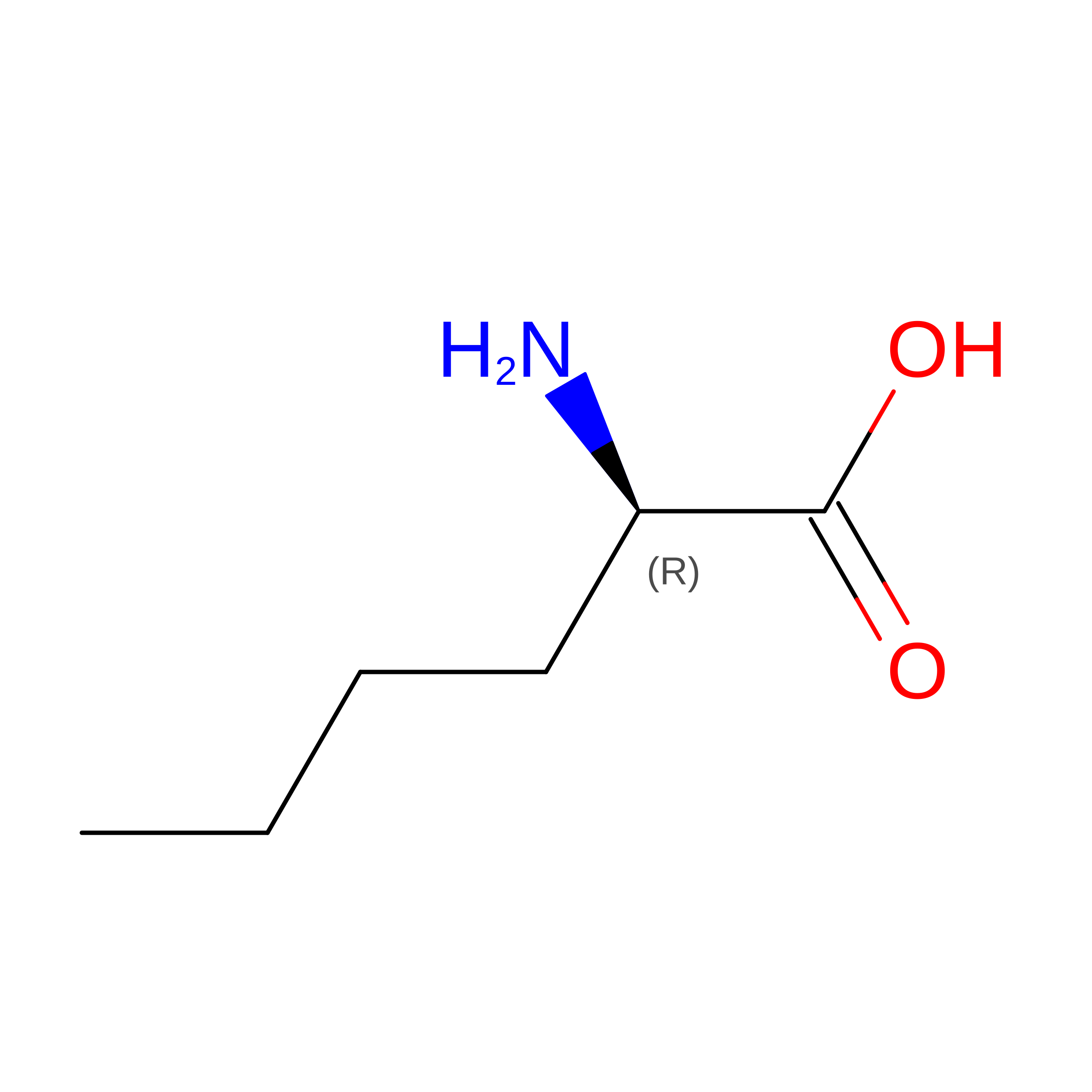
D-PEPTIDE LINKINGC6 H13 N O2Not Available
DLY
Query on DLY
C, D
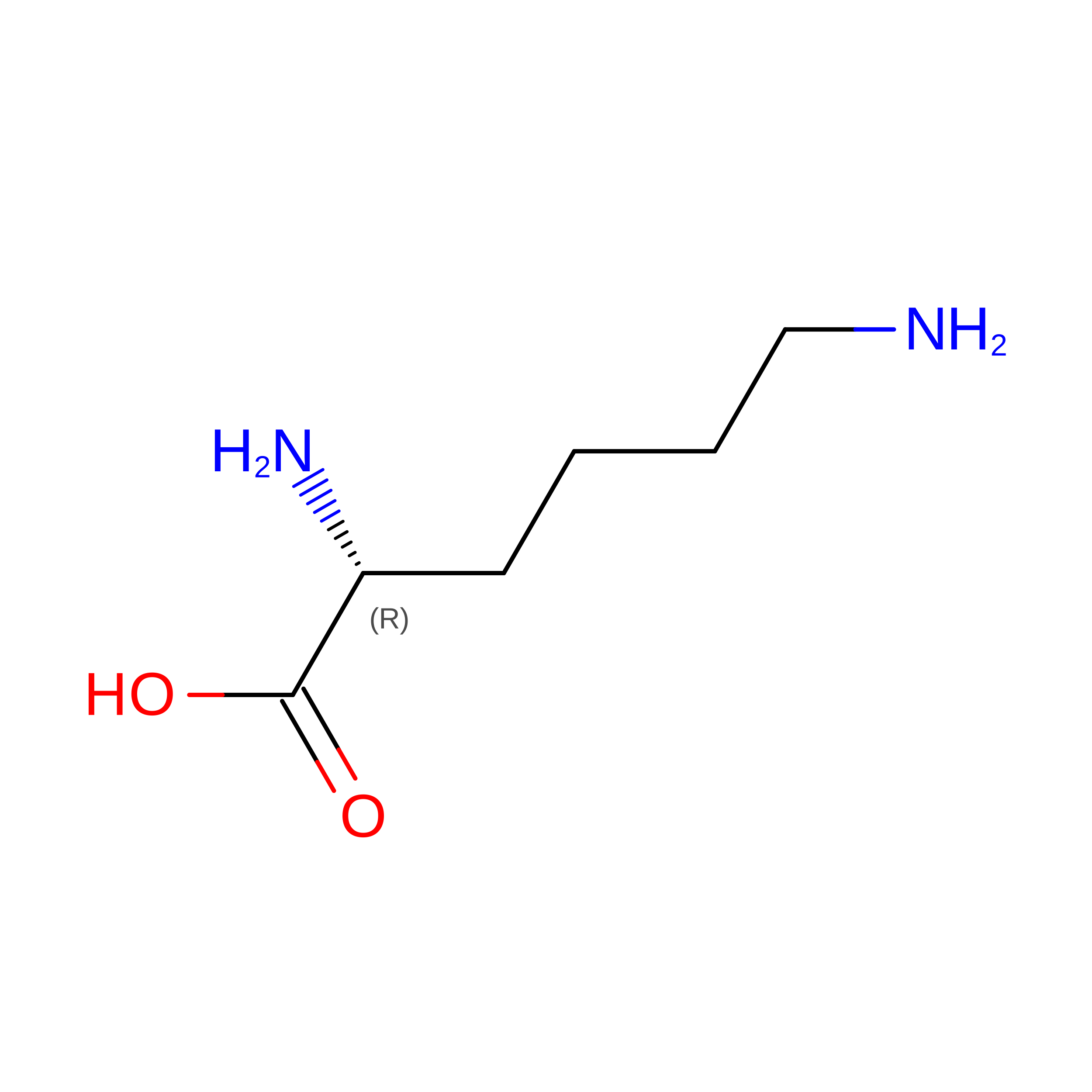
D-PEPTIDE LINKINGC6 H14 N2 O2Not Available
DNE
Query on DNE
C, D
D-PEPTIDE LINKINGC6 H13 N O2Not Available
DPN
Query on DPN
C, D
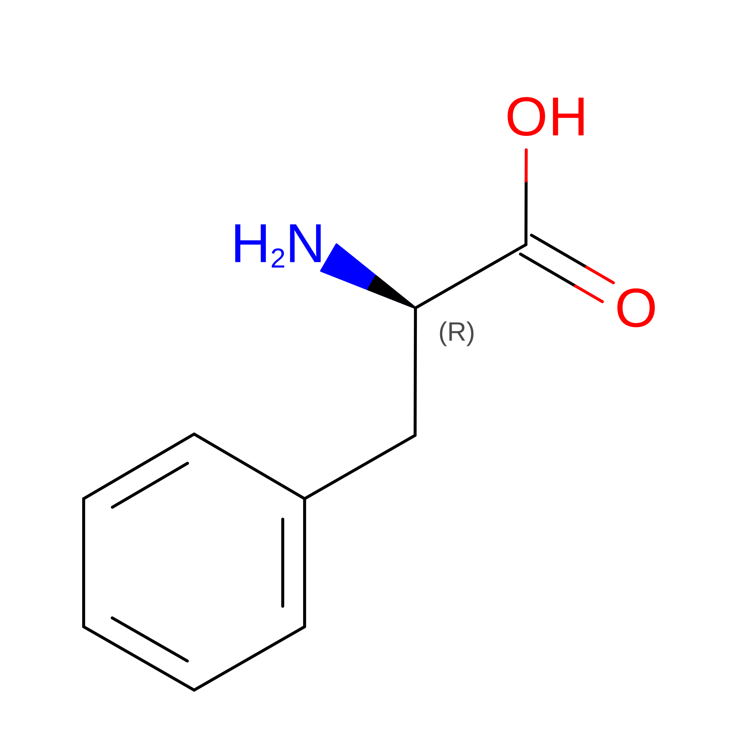
D-PEPTIDE LINKINGC9 H11 N O2Not Available
DPR
Query on DPR
C, D
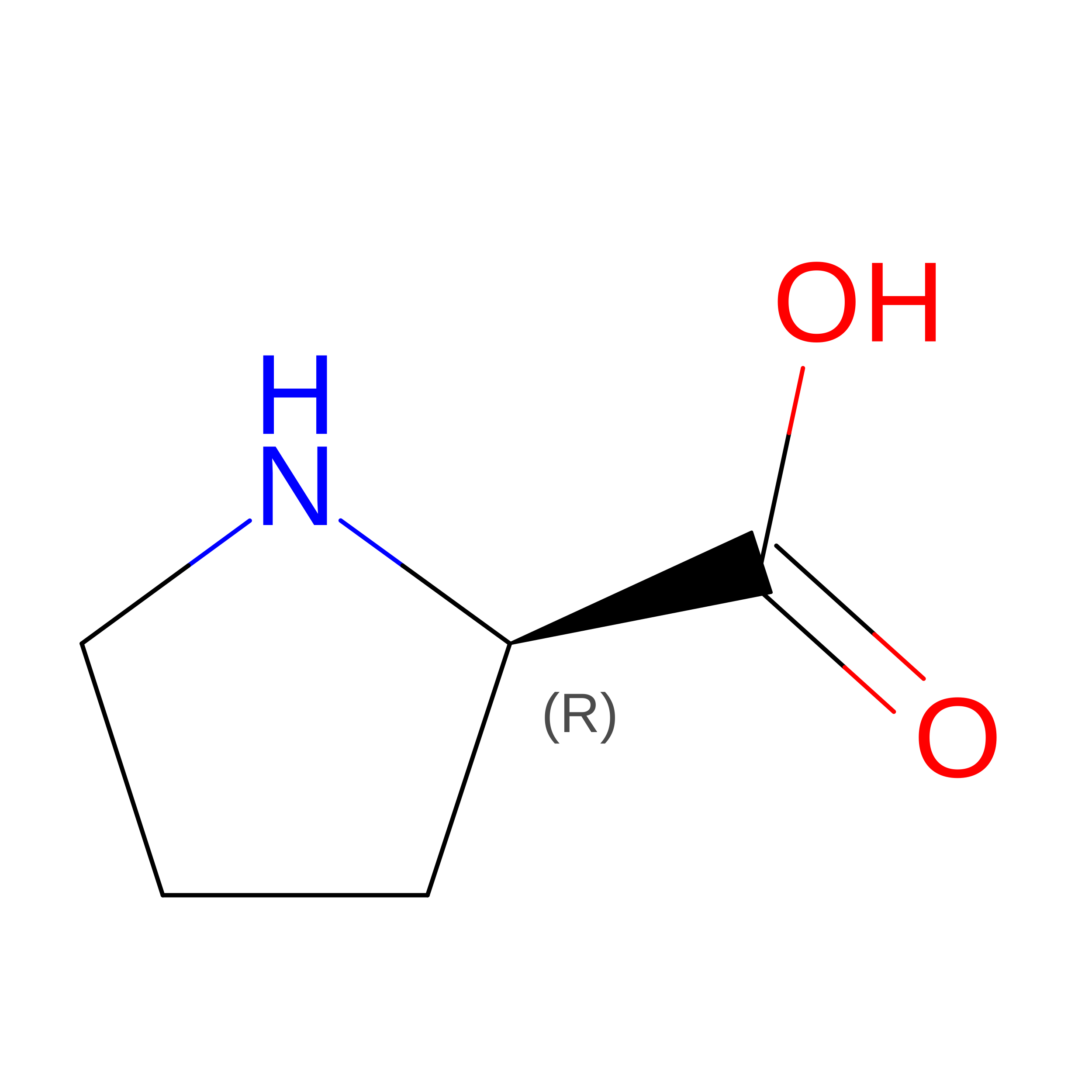
D-PEPTIDE LINKINGC5 H9 N O2Not Available
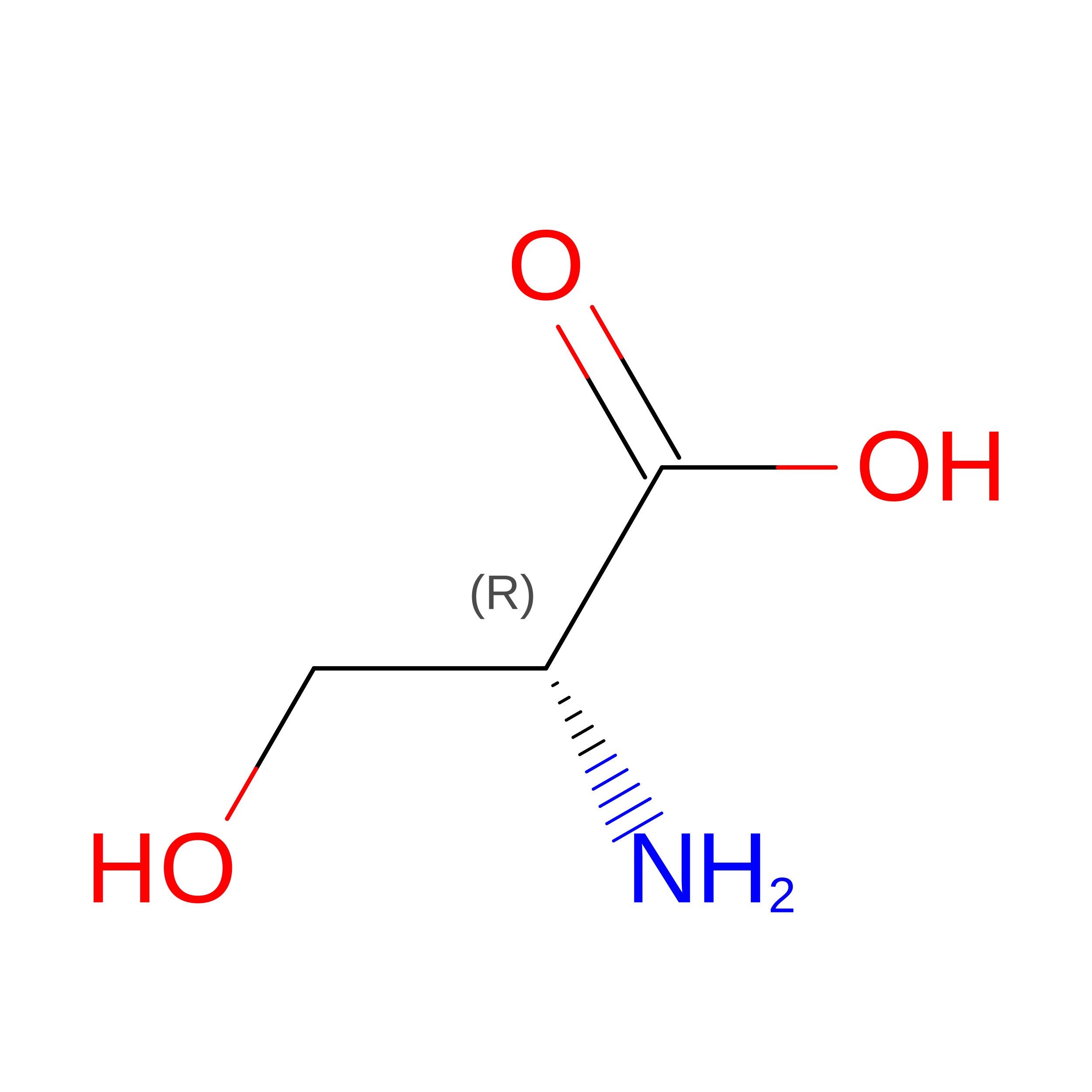
DSG
Query on DSG
C, D
D-PEPTIDE LINKINGC4 H8 N2 O3Not Available
DSN
Query on DSN
C, D
D-PEPTIDE LINKINGC3 H7 N O3Not Available
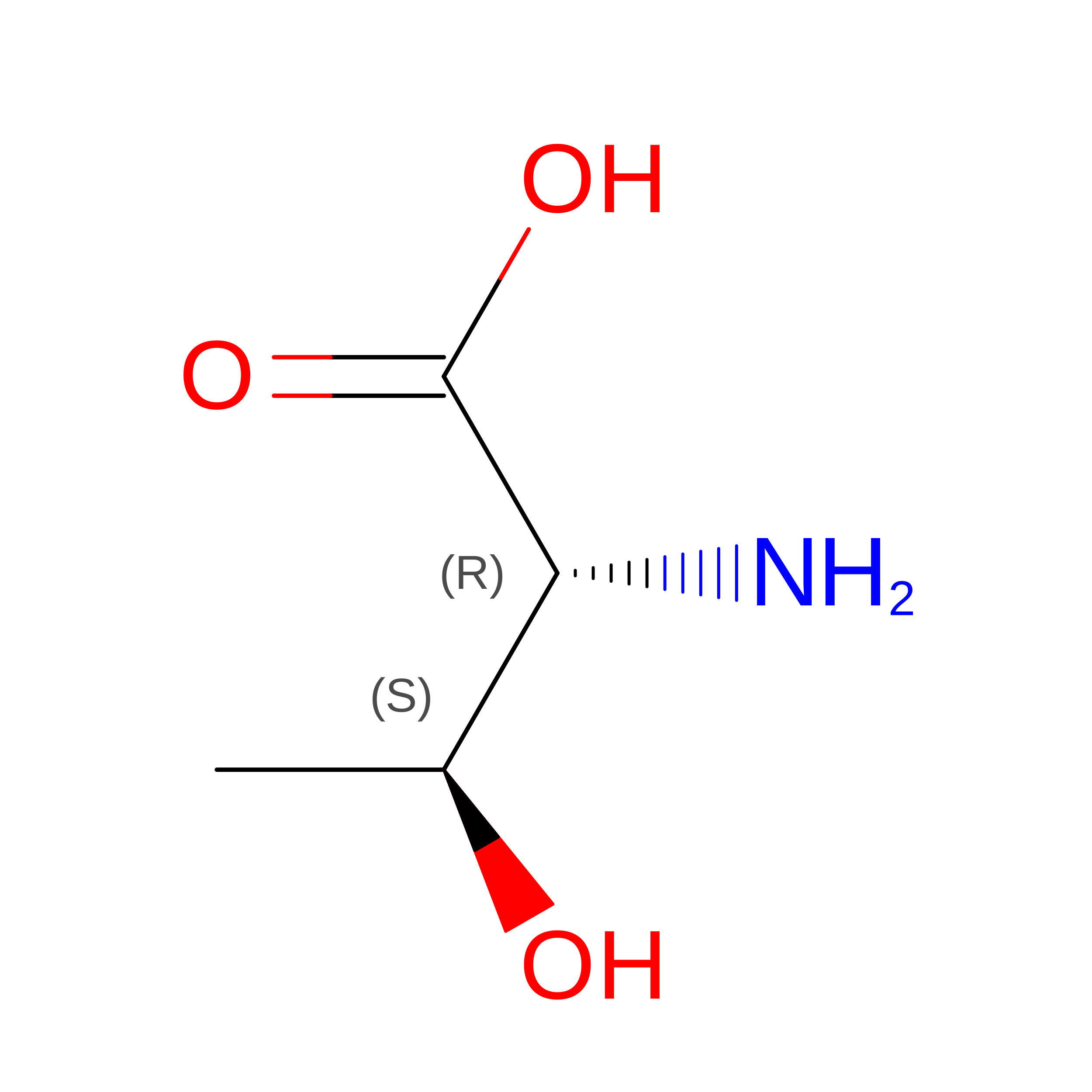
DTH
Query on DTH
C, D
D-PEPTIDE LINKINGC4 H9 N O3Not Available
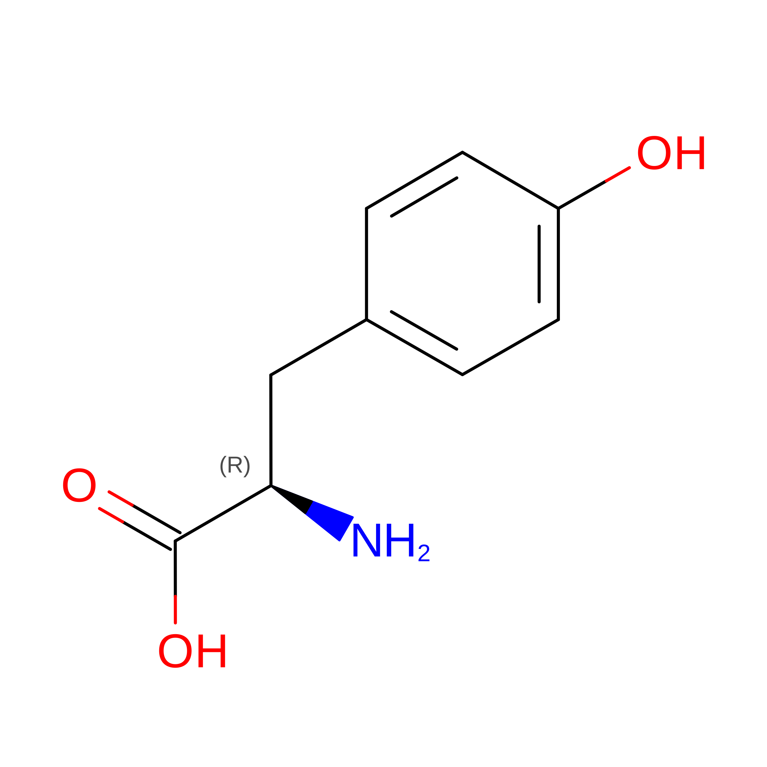
DTY
Query on DTY
C, D
D-PEPTIDE LINKINGC9 H11 N O3Not Available
DVA
Query on DVA
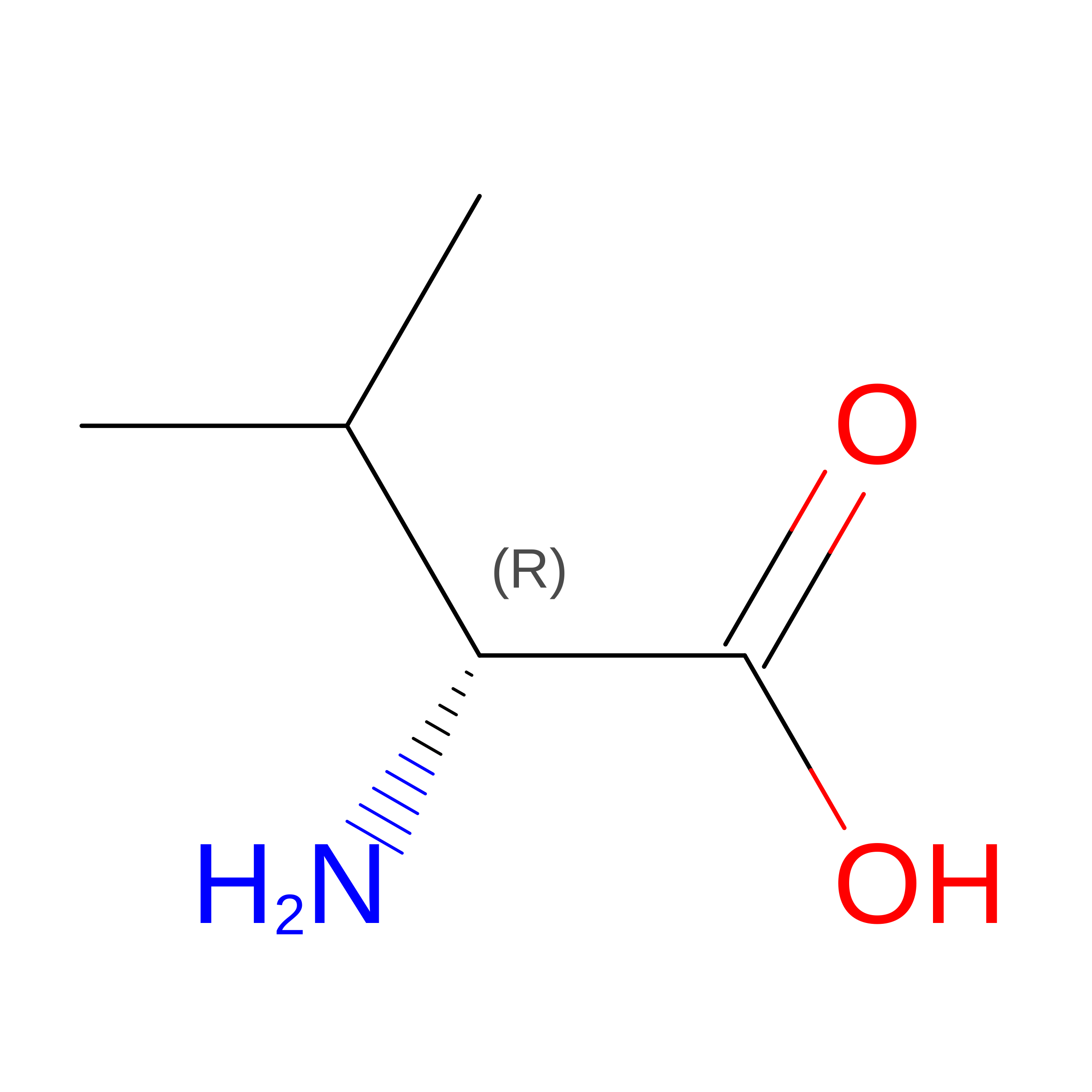
C, D
D-PEPTIDE LINKINGC5 H11 N O2Not Available

Experimental Data & Validation

Experimental Data

  • Method: X-RAY DIFFRACTION
  • Resolution: 1.15 Å
  • R-Value Free:  0.222 (Depositor), 0.222 (DCC) 
  • R-Value Work:  0.202 (Depositor) 
  • R-Value Observed: 0.203 (Depositor) 
Space Group: P 1
Unit Cell:
Length ( Å )Angle ( ˚ )
a = 38.24α = 98.15
b = 38.26β = 98.15
c = 48γ = 109.67
Software Package:
Software NamePurpose
PHENIXrefinement

Structure Validation

View Full Validation Report


Entry History 

Deposition Data

Revision History  (Full details and data files)

  • Version 1.0: 2016-11-02
    Type: Initial release
  • Version 1.1: 2016-11-16
    Changes: Database references
  • Version 1.2: 2022-02-09
    Changes: Database references, Derived calculations
  • Version 1.3: 2023-11-08
    Changes: Data collection, Refinement description
  • Version 1.4: 2023-11-15
    Changes: Data collection