Active site gate of M32 carboxypeptidases illuminated by crystal structure and molecular dynamics simulations
Sharma, B., Jamdar, S.N., Ghosh, B., Yadav, P., Kumar, A., Kundu, S., Goyal, V.D., Makde, R.D.(2017) Biochim Biophys Acta 1865: 1406-1415
- PubMed: 28844748 Search on PubMed
- DOI: https://doi.org/10.1016/j.bbapap.2017.07.023
- Primary Citation Related Structures:
5GIV - PubMed Abstract:
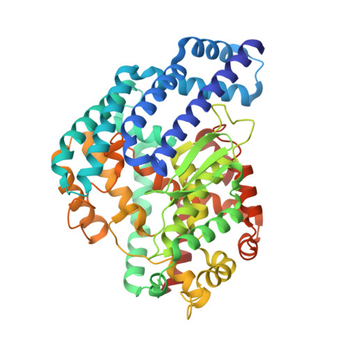
Enzyme gates are important dynamic features that regulate function. Study of these features is critical for understanding of enzyme mechanism. In this study, the active-site gate of M32 carboxypeptidases (M32CP) is illuminated. Only a handful of members of this family have been structurally and functionally characterized and various aspects of their activity and mechanism are yet not clarified. Here, crystal structure of putative M32CP from Deinococcus radiodurans (M32dr) was solved to 2.4Å resolution. Enzymatic assays confirmed its identity as a carboxypeptidase. Open and relatively closed conformations observed in the structure provided supporting evidence for previously hypothesized hinge motion in this family of enzymes. Molecular dynamics simulations of 1.5μs displayed distinct open and closed conformations revealing amplitude of the motion to be beyond what was observed in the crystal structure. Hinge region and anchoring region of this shell-type gate were identified. A small displacement of 3Å and a helical tilt of 9° propagated by the hinge region translates into a 10Å motion at the top of the gate. The dynamics of the gate was supported by our mutagenesis experiment involving formation of disulphide bond across helices of the gate. The nearly inactive mutant enzyme showed 65-fold increase in the enzymatic activity in presence of reducing agent. Further, while a previously proposed structural basis would have led to its classification in subfamily II, experimentally observed substrate length restriction places M32dr in subfamily I of M32CPs.
- High Pressure and Synchrotron Radiation Physics Division, Bhabha Atomic Research Centre, Mumbai, India; Department of Biochemistry, University of Delhi South Campus, New Delhi, India.
Organizational Affiliation: